Myelographie


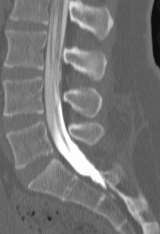


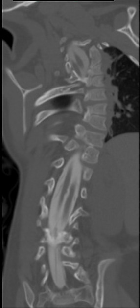
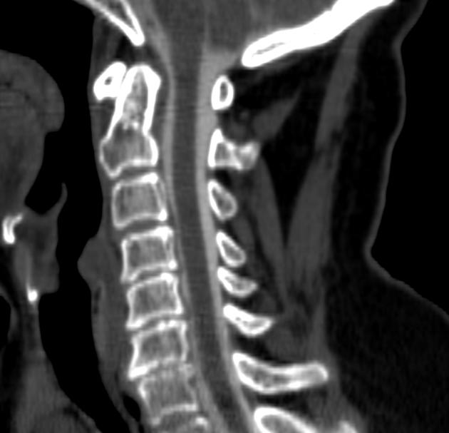
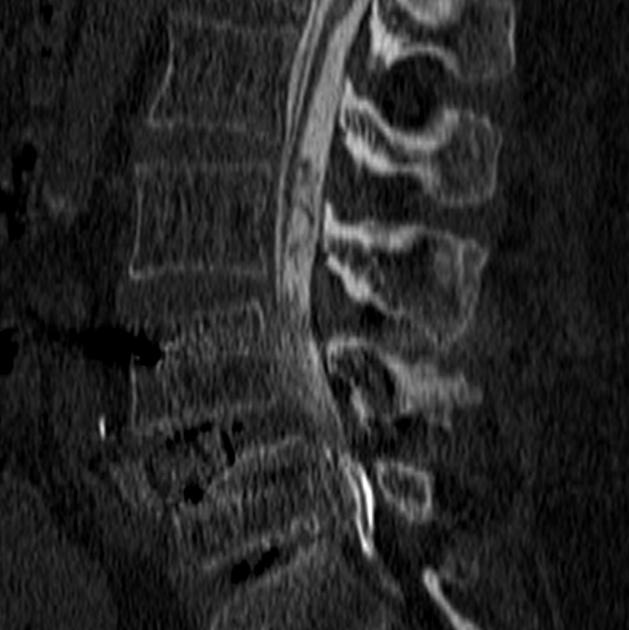
Myelography is a form of imaging intended to evaluate the subarachnoid spaces within the spinal canal. It is now usually performed with either CT or MR imaging of the spine after injection of an intrathecal iodinated or gadolinium-based contrast agent. MR myelography may also provide evaluation without the need for intrathecal gadolinium by utilizing specific fluid-sensitive acquisition techniques.
Although conventional (i.e. fluoroscopic) myelography has been largely supplanted by modern CT and (especially) MRI of the spine, it remains a useful tool in the evaluation of spondylosis. CT myelography is especially useful in those patients who cannot undergo MRI.
History
The concept of using a contrast agent to better image the contents of the spinal canal originated as an extension of pneumoencephalography, wherein air was instilled into the subarachnoid space to provide "negative" contrast for evaluating structures of the brain. Although crude, requiring extraction of an equivalent volume of cerebrospinal fluid to instilled air, and extremely painful, air-contrast myelography (or pneumomyelography) techniques allowed some visualization of the intraspinal soft tissues as imaged by fluoroscopy.
In the 1920 and 1930s, myelography using 'positive' liquid contrast agents became more common. Initial agents included oil-based compounds such as Lipiodol, and water-soluble compounds, such as Thorotrast, and Abrodil.
Iophendylate (Pantopaque) was a popular myelography agent until the 1970s. An oil-based agent, it was not readily reabsorbed and thus often remained trapped within the subarachnoid spaces for decades . As with many of its pharmacological predecessors, it was notorious for causing arachnoiditis. It was withdrawn from clinical use in 1988 .
Siehe auch:


 Assoziationen und Differentialdiagnosen zu Myelographie: