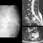
Aneurysmatische Knochenzyste der Wirbelsäule

Pitfalls in
the diagnosis of common benign bone tumours in children. Aneurysmal bone cyst of the second cervical vertebra (C2): a Lateral X-ray of the cervical spine: large expansile lytic lesion of the posterior arch C2 (between arrowheads). b CT, axial slice, bone windowing: confirmation of the lesion, with extreme thinning and focal interruption of the bone cortex. c CT, axial slice, soft-tissue windowing: fluid-fluid levels (arrows) are identified inside the cyst

Spinal
osteoblastoma: a retrospective study of 35 patients’ imaging findings with an emphasis on MRI. A 28-year-old man with aneurysmal bone cyst (ABC). A 28-year-old man with a lesion on the right accessory. The nidus can be visualized well on axial CT (a, b), especially in the bone window (b). The nidus exhibits isointense signal on T1WI (c) and hyperintense signal on FS T2WI (d). ABC appears conspicuous on MRI (e, f), with a typical finding of the fluid–fluid level (f)

Aneurysmal
bone cyst • Aneurysmal bone cyst T11 - Ganzer Fall bei Radiopaedia

Aneurysmal
bone cyst • Aneurysmal bone cyst - Ganzer Fall bei Radiopaedia

Aneurysmal
bone cyst • Aneurysmal bone cyst - cervical spine - Ganzer Fall bei Radiopaedia

Aneurysmal
bone cyst • Vertebral aneurysmal bone cyst - Ganzer Fall bei Radiopaedia

Aneurysmal
bone cyst • Aneurysmal bone cyst - sacrum - Ganzer Fall bei Radiopaedia

Sacral
lesions • Giant cell tumor with aneurysmal bone cyst of sacrum - Ganzer Fall bei Radiopaedia

School ager
with lower back and left buttock pain and a limpLateral radiograph of the lumbar spine shows an expansile lesion in the S2 vertebral body. Axial and sagittal T2 MRI of the lumbar spine shows the S2 expansile lesion to be well defined and to have fluid-fluid levels within it.The diagnosis was aneurysmal bone cyst of the sacrum.

Aneurysmal
bone cyst • Aneurysmal bone cyst - Ganzer Fall bei Radiopaedia

Bone up on
spinal osseous lesions: a case review series. Aneurysmal bone cyst. Lateral radiograph (a) of the cervical spine demonstrates a large, osteolytic, expansile mass arising from the C2 vertebral body, resulting in significant bony remodeling. The mass is sharply defined and demonstrates thin sclerotic margins, also known as an “eggshell cortex” (orange arrows). Sagittal CT image of the cervical spine (b) confirms the lesion within the C2 vertebral body. Axial CT image in the soft tissue window (c) shows fluid-fluid levels (orange arrows), compatible with hemorrhage. Axial CT image in the bone window (d) shows near complete obliteration of the posterior arch with some areas of remnant thin sclerotic rim, "eggshell cortex" (white arrow)

Bone up on
spinal osseous lesions: a case review series. Aneurysmal bone cyst. MRI imaging in the same patient as Fig 9. Pre-contrast axial T1-weighted image (a) confirms fluid-fluid levels within the lesion (orange arrows), indicating hemorrhage with sedimentation of varying stages of blood products. Post-contrast axial T1-weighted image (b) demonstrates enhancement of the septa within the lesion (orange arrows). Note a partially hypointense rim, representing cortex (white arrow). Axial T2-weighted image (c) re-demonstrates a destructive lesion with fluid-fluid levels (orange arrows); varying signal intensity corresponds to differing stages of blood product degradation

School ager
with back pain and incontinence who has been unable to walk for several weeks. Sagittal (above left) and axial (above right) CT without contrast of the cervical spine shows an expansile lytic lesion with a thin margin in the spinous and transverse processes of the T1 vertebral body. There is marked anterolisthesis of T1 on T2. Sagittal T2 MRI without contrast of the cervical spine (below left) shows the anterolisthesis at T1 and T2 causing extreme cord compression while an axial T2 MRI obtained at this level (below right) shows a fluid-fluid level in the lesion to the anterior and right of the vertebral body.The diagnosis was aneurysmal bone cyst of the spine.
Aneurysmatische Knochenzyste der Wirbelsäule
Siehe auch:

 Assoziationen und Differentialdiagnosen zu Aneurysmatische Knochenzyste der Wirbelsäule:

