Varizen des Ligamentum teres uteri

Varicocele
des Ligamentum teres uteri war auffällig als tastbar, weiche Vorwölbung in der Leiste bei einer Schwangeren. Kein Nachweis einer Hernierung.
A 28-year-old
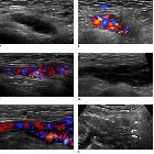
women at the 21st week of pregnancy with round ligament varicosities. A. Grayscale transverse ultrasonography shows an ovoid multiseptated cystic mass in the left groin. B. Color Doppler ultrasonography shows that the lesion is hypervascular. After 5 weeks, follow-up ultrasonography was performed (C-F). The mass in the left groin shows slightly enlarged and composed of multiple anechoic serpentine tubular channels on grayscale ultrasonography (not shown). C. Color Doppler sagittal ultrasonography during Valsalva maneuver. The mass expands and shows marked flow augmentation. D, E. Sagittal ultrasonography of the left groin through the inguinal canal in an erect position. The lesion is enlarged and the vascularities of the mass are markedly engorged. F. These varicose veins between the markers continue to the left parauterine space (arrows) through the inguinal canal (arrowheads).

Varicosis am
Ligamentum teres uteri in der Schwangerschaft (32. Schwangerschaftswoche). Es zeigen sich varikös veränderte, spongiforme Gefäßkonvolute im Leistenkanal bis an die V. femoralis heranreichend. Im Farbdoppler waren kräftige Flusssignale darstellbar. Der Befund darf nicht mit einer Leistenhernie verwechselt werden.
Varizen des Ligamentum teres uteri treten klinisch als weiche Schwellung in der Leiste insbesondere bei Schwangeren ab dem zweiten Trimenon in Erscheinung. Sie dürften nicht mit einer Inguinalhernie verwechselt werden, zumal sich beide Pathologien im Pressversuch vergrößern. Die Unterscheidung gelingt zuverlässig mit Einsatz der Dopplersonographie, in der sich kräftige Flusssignale in den tubulär-spongiform erweiterten Gefäßen vom Leistenkanal Richtung Labia majora nachweisen lassen. Diese Drainagevenen des Ligamentum teres uteri münden in die Vena epigastrica inferior.
Siehe auch:


 Assoziationen und Differentialdiagnosen zu Varizen des Ligamentum teres uteri:

