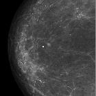
Brustdichte in der Mammographie


Breast density refers to the amount of fibroglandular tissue in a breast relative to fat. It can significantly vary between individuals and within individuals over a lifetime.
Classification
There are four descriptors for breast density on mammography in the 5 edition of BI-RADS :
- a: the breasts are almost entirely fatty
- b: there are scattered areas of fibroglandular density
- c: the breasts are heterogeneously dense, which may obscure small masses
- d: the breasts are extremely dense, which lowers the sensitivity of mammography
Compared to the previous edition, the estimation of dense tissue percentages were replaced by a description of subjectively estimated breast density associated with changes in the sensitivity of mammography, since the effect of percentage breast density as an indicator for breast cancer risk takes on a greater importance compared to the masking effect of dense fibroglandular tissue on mammographic depiction of noncalcified lesions .
An optional description of the location of densities can be included as a second sentence for the scattered and heterogeneous categories. The radiologist is encouraged to assign a category based on the concern that areas of dense fibroglandular breast tissue could mask underlying cancer .
There are analogous BI-RADS categories for the amount of fibroglandular tissue on breast MRI:
- almost entirely fat
- scattered fibroglandular tissue
- heterogeneous fibroglandular tissue
- extreme fibroglandular tissue
Breast density assessment is qualitative and subject to interobserver variability .
For other classifications of historical significance, see the article on parenchymal patterns in breast imaging.
Significance
In dense breasts, the incidence of invasive ductal carcinoma is higher than in other groups of breast density, likely as a combination of the amount of gland present as well as possible observation error due to the lower sensitivity of detection.
The use of density determination is now mandatory in some states in the United States, and patients must be informed of the fact that they have "dense" breasts, which usually is defined as heterogeneously dense or extremely dense.
In dense breasts, screening ultrasound is potentially beneficial. Contrast-enhanced MRI is also advocated as an adjunct to screening mammography in extremely dense breasts; however, no large data is available in this regard. The DENSE Trial is undergoing to evaluate MRI as an additional screening tool in women aged 50-75 years with extremely dense breasts .
Factors that can increase breast density
- hormone replacement therapy: the effect is greater with combination hormone therapy than with estrogen therapy alone
- pregnancy
- lactation
- weight loss (from a reduction of breast fat)
- breast cancer: especially inflammatory breast cancer
- inflammation: mastitis
Factors that can decrease breast density
- age, postmenopausal state
- medications, e.g. danazol
- vitamin D and calcium intake in pre-menopausal women
- increasing age
- weight gain
- acromegaly
Siehe auch:
- Breast imaging-reporting and data system (BI-RADS)
- Mastitis
- inflammatorisches Mammakarzinom
- altered breast density between two mammograms
- Neoplasien der Mamma
- Boyd classification mammography
- Wolfe classification of breast density
- low weight effect on mammogram
und weiter:

 Assoziationen und Differentialdiagnosen zu Brustdichte in der Mammographie:


