double chamber right ventricle (DCRV)
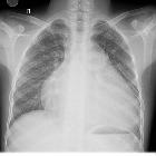
Double
Trouble. Chest X-ray of the patient shows gross cardiomegaly.

Double
Trouble. CT angiography MPR image displays the thin hypodense band in the left ventricular outflow tract consistent with subaortic membrane. LV: Left ventricle.

Double
Trouble. CT angiography axial image displays the thin hypodense band in the left ventricular outflow tract consistent with subaortic membrane

Double
Trouble. CTA axial image displays the thick muscle bundle (arrow) in the right ventricle outflow tract.

Double
Trouble. Reformatted MPR image displays the thick muscle bundle (arrow) in the right ventricle outflow tract. RV: right ventricle; LV: Left ventricle; PA: Main pulmonary artery.
 nicht verwechseln mit: double outlet right ventricle (DORV)
double chamber right ventricle (DCRV)
Siehe auch:


 Assoziationen und Differentialdiagnosen zu double chamber right ventricle (DCRV):
