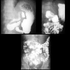
Volvulus des Colon transversum

Transverse
colon volvulus: Case report and review of the literature. AP abdominal plain radiograph. Low-grade obstructive ileus: marked dilation of colon and small bowel loops. Faecal content in sigma is detected.

Transverse
colon volvulus: Case report and review of the literature. Volumetric reconstruction in AP coronal projection. Colonic dilation with change of calibre in transverse colon (arrow).

Transverse
colon volvulus: Case report and review of the literature. Contrast-enhanced CT in portal phase, axial projection. Colonic distention with collapsed descending colon (arrowhead).

Transverse
colon volvulus: Case report and review of the literature. Contrast-enhanced CT in portal phase, coronal projection. Colonic dilation with collapsed descending colon (arrowheads). Sudden change of calibre in transverse colon (white arrow).

Transverse
colon volvulus: Case report and review of the literature. Contrast-enhanced CT in portal phase, coronal projection. ‘Whirlpool sign’. At the point of calibre change, twirling of transverse mesocolon vessels is seen (black arrowhead) shaping the classic ‘whirl’ image.

Transverse
colon volvulus: Case report and review of the literature. Contrast-enhanced CT in portal phase, sagittal projection. ‘Bird’s beak sign’: Transverse colon dilation with sudden sharpness and change of calibre.
Volvulus des Colon transversum
Siehe auch:

 Assoziationen und Differentialdiagnosen zu Volvulus des Colon transversum:

