posttraumatische Lipoatrophie
Imaging
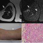
spectrum of abnormal subcutaneous and visceral fat distribution. A 59-year-old woman with post blunt trauma lipoatrophy on the right upper arm. a Axial T2-weighted and (b) STIR images show thinning of subcutaneous fat on the lateral aspects (arrows) with multiple high signal nodules (small arrows) showing small peripheral fat signal areas. c Photograph shows thinning of the lateral aspect of the upper arm with small hump (arrow). d Histologically fat necrosis with lipogranuloma was proven. Variably sized lipid vacuoles are surrounded by foam cells, foreign body-type (arrows), and Touton giant cells (arrowhead) in the resected lipogranuloma. A adipocytes, F fibrosis (hematoxylin-eosin stain, × 100)
Imaging
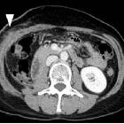
spectrum of abnormal subcutaneous and visceral fat distribution. A 59-year-old woman with post-surgery lipoatrophy. a CT image before operation shows calcified soft tissue density mass anterior to the right kidney (arrow). Right nephrectomy was done, and the lesion was pathologically diagnosed as dedifferentiated liposarcoma. b CT image one month after surgery shows fluffy opacity in the subcutaneous fat around the operated area (arrowhead) with abdominal wall muscle swelling. c CT image after 8 years shows local lipoatrophy (double arrow) with muscle atrophy


 Assoziationen und Differentialdiagnosen zu posttraumatische Lipoatrophie: