epidural abscess

Paediatric
acute mastoiditis complicated by Bezold abscess and epidural empyema without bone erosion. Axial contrast-enhanced CT of the head demonstrates filling defect with surrounding enhancement in the left sigmoid sinus and Bezold abscess.

Paediatric
acute mastoiditis complicated by Bezold abscess and epidural empyema without bone erosion. Coronal contrast-enhanced CT of the head demonstrates filling defect with surrounding enhancement in the left sigmoid sinus and Bezold abscess.

Paediatric
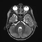
acute mastoiditis complicated by Bezold abscess and epidural empyema without bone erosion. Absent flow void in the left sigmoid sinus with area of high T2 signal lateral to the sinus

Paediatric
acute mastoiditis complicated by Bezold abscess and epidural empyema without bone erosion. DWI (b0): Area of restricted diffusion (relative to CSF) lateral to the left sigmoid sinus suggestive of an extra-axial empyema

Paediatric
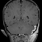
acute mastoiditis complicated by Bezold abscess and epidural empyema without bone erosion. Bezold abscess lateral to the mastoid (arrow) with surrounding enhancement of the sternocleidomastoid muscle. Enhancement around the sigmoid sinus suggestive of thrombosis and/or intracranial collection (asterisk)

Paediatric
acute mastoiditis complicated by Bezold abscess and epidural empyema without bone erosion. There is swelling and enhancement of the left sternocleidomastoid muscle (black arrowhead). Note the normal right stenocleidomastoid muscle (white arrow)

A rare
case as different cause of retropharyngeal and spinal epidural abscess: spondylodiscitis. Axial and sagittal sections of CT: the stars show the retropharyngeal abscess, and the arrows show the degeneration of C5–6 vertebrae

A rare
case as different cause of retropharyngeal and spinal epidural abscess: spondylodiscitis. T2-weighted, sagittal section of vertebra MRI: the star shows the retropharyngeal abscess, and the arrow show the spinal epidural abscess. Myelopathic signal changes of the spinal cord are clearly seen

Brucellosis
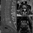
presenting as a spinal epidural abscess in a 41-year-old farmer: a case report. A sagittal pre-contrast T1-weighted spin-echo (SE) image shows a fluid collection in the posterior epidural space with low signal intensity, that is anteriorly compressing the dorsal sac and decreased signal intensity of the L5 and S1 vertebral bodies.
 Assoziationen und Differentialdiagnosen zu epiduraler Abszess:


