idiopathisches akutes Skrotalödem

The
“fountain sign” in a case of idiopathic scrotal oedema, combined with cryptorchidism. Transverse power Doppler image showing thickened, hypoechogenic and hypervascularized scrotal wall.

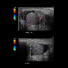
The
“fountain sign” in a case of idiopathic scrotal oedema, combined with cryptorchidism. Transverse colour Doppler image of the scrotum demonstrating increased blood flow signals within the peritesticular scrotal soft tissues. The signal pattern resembles a coloured fountain and is created by the intense hyperaemia (arrow).

The
“fountain sign” in a case of idiopathic scrotal oedema, combined with cryptorchidism. Schematic representation of the fountain sign.


 Assoziationen und Differentialdiagnosen zu idiopathisches akutes Skrotalödem: