Ureterklappen
Double
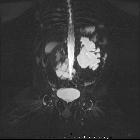
trouble: diagnostic dilemma in a rare association of proximal ureteric valve with orthotopic ureterocele—case report. Coronal view of magnetic resonance urogram showing left gross hydronephrosis with cutoff at pelviureteric junction

School ager
with chronic left flank pain. Sagittal US of the left kidney (above) shows a moderate amount of hydronephrosis. Image from the excretory phase of an intravenous pyelogram exam (below left) shows a normal right renal collecting system and a markedly dilated left renal collecting system and proximal left ureter. Image from a retrograde ureteroscopy exam (below right) shows normal caliber of the distal and middle left ureter with a sharp area of narrowing in the proximal left ureter with a markedly dilated proximal left ureter above it.The diagnosis was a left ureteral valve.
 nicht verwechseln mit: Urethralklappe

 Assoziationen und Differentialdiagnosen zu Ureterklappen:
