schwannoma of the hypoglossal nerve

Imaging of
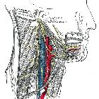
cranial nerves: a pictorial overview. Hypoglossal schwannoma. MRI T2-weighted axial image (a), T1-weighted post-gadolinium axial (b), and coronal images (c) demonstrate an extra-axial expansive lesion (dotted circles), mildly hyperintense with heterogenous contrast enhancement, surrounded by cystic components. The mass is located along the course of the right hypoglossal nerve. CT (bone window, d) clearly shows the enlargement of the right hypoglossal canal (arrow). Atrophy of the right tongue muscles (asterisks) is well visible at CT (e) and T1-weighted axial sequence (f) as hypodense and hyperintense area, respectively


 Assoziationen und Differentialdiagnosen zu schwannoma of the hypoglossal nerve: