lytische Läsionen des Kalkaneus


Pseudotumor
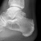
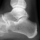
des Kalkaneus: An typischer Stelle zeigt sich eine zystoid imponierende Rarefizierung der Spongiosa Struktur im Calcaneus. Differenzialdiagnostisch wäre ein Calcaneus Lipom denkbar, dieses hat jedoch häufig eine zentrale Verkalkung.


Intraossäres
Lipom im Kalkaneus bei einer 36-jährigen Frau. Typisches Bild mit zentraler Verkalkung.

Unicentric
epithelioid hemangioendothelioma of the calcaneus: a case report and review of literature. Pre-operative radiograph of large lytic lesion in the calcaneus

Unicentric
epithelioid hemangioendothelioma of the calcaneus: a case report and review of literature. Preoperative sagittal T2 image with some heterogeneity within the lesion surrounding bony edema, sclerotic border around the lesion, but with concern for cortical breakthrough in the subtalar joint

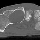
Unicentric
epithelioid hemangioendothelioma of the calcaneus: a case report and review of literature. Axial CT scan of lesion preoperatively displaying concern for cortical destruction

Distribution
patterns of foot and ankle tumors: a university tumor institute experience. Osteolytic lesions of the calcaneus with different radiographic appearance and varying aggressive behaviour: (a) Ewing sarcoma in a 31-year old male patient, (b) simple (calcaneal) bone cyst in a 11-year old male patient, (c) secondary squamous cell carcinoma based on chronic osteomyelitis in a 82-year old male patient and (d) low-grade chondrosarcoma in a 45-year old female

Rare case of
pediatric calcaneal osteosarcoma masquerading as a cystic lesion. Axial bone and soft tissue window, sagittal bone window and coronal bone window CT of the ankle demonstrate a permeative soft tissue lesion in the calcaneous without invasion of the physis/epiphysis with expansion of the bone and cortical breakthrough without ossification. Increased linear lace like density in the lesion suggests chondroid or ossific matrix

Cementoma of
the calcaneus: a case report. Lateral radiograph of the left calcaneum. An expansive, partially osteolytic lesion is displayed, with well-defined margins, and an eccentric calcified matrix

Cementoma of
the calcaneus: a case report. Computed tomography (CT) scans of the calcaneum. a Saggital reconstruction image and (b) coronal reconstruction image show an osteolytic lesion with sclerotic margin and an eccentric focus of matrix calcification adjacent to soft tissue dense with an arc-shaped fat band
 Assoziationen und Differentialdiagnosen zu lytische Läsionen des Kalkaneus:


