carpal tunnel


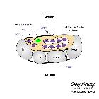
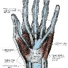
The carpal tunnel is a fibro-osseous canal in the anterior (volar) wrist that acts as a passageway for structures between the forearm and the anterior hand.
Gross anatomy
Boundaries
- superficial border (roof): flexor retinaculum
- deep border (floor): carpal groove (formed by palmar aspect of carpal bones)
- medial/ulnar border: lateral surface of hamate
- lateral/radial border: medial surface of trapezium
Attachments
- medial/ulnar: pisiform bone and hook of hamate
- lateral/radial: scaphoid tubercle and ridge of trapezium
Contents
The carpal tunnel contains the following structures, from superficial to deep:
- flexor digitorum superficialis tendons (four) (with middle and ring finger more superficial to the index and little finger)
- median nerve (laterally)
- flexor pollicis longus tendon (laterally)
- flexor digitorum profundus tendons (four)
Note, the flexor carpi radialis is often wrongly stated to be within the carpal tunnel. Its course is in fact between the superficial and deep layers of the flexor retinaculum itself within its own compartment.
The tendons within the carpal tunnel are surrounded by synovial sheaths: one which envelops the flexor digitorum superficialis and profundus together, and a separate sheath for the flexor pollicis longus.
Onto the surface of the medial aspect is formed the Guyon's canal through which runs the ulnar nerve and artery (ulnar nerve more medial), superficial to the tunnel. More laterally, the palmaris longus is attached to the superficial surface. The palmar cutaneous branch of the median nerve runs superficial.
Variant anatomy
Variant anatomical structures within the carpal tunnel :
- persistent median artery of the forearm
- bifid median nerve
- presence of the motor branch of the median nerve within the carpal tunnel
- presence of palmaris longus tendon within the carpal tunnel
Related pathology
Siehe auch:
und weiter:

 Assoziationen und Differentialdiagnosen zu Karpaltunnel:
