Buschke-Löwenstein-Tumor

Successful
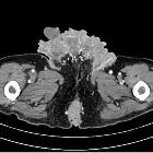
treatment of paraneoplastic hypercalcemia in a patient with giant condyloma acuminatum: a case report. Extract of pelvis computed tomography scan showing a destructive, polycyclic growing tumor. This is a picture of a computed tomography scan that we made to exclude bone metastasis. It shows the tumor mass, growing from the anal region (below in the picture) to the groin region (above). Furthermore it shows the local destructive grow-pattern.
An
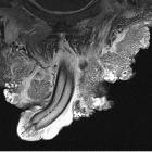
extraordinary case of Buschke–Lowenstein tumor: multiple localization, malignant transformation, and clinical insights—a case presentation and literature review. CT image of the inguinal lesion. CT image was done for further investigation of hypercalcemia
An
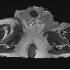
extraordinary case of Buschke–Lowenstein tumor: multiple localization, malignant transformation, and clinical insights—a case presentation and literature review. MRI image of the inguinal and penile lesions
An
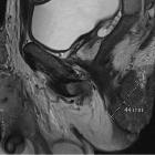
extraordinary case of Buschke–Lowenstein tumor: multiple localization, malignant transformation, and clinical insights—a case presentation and literature review. MRI image of the perineal and inguinal lesions
An
extraordinary case of Buschke–Lowenstein tumor: multiple localization, malignant transformation, and clinical insights—a case presentation and literature review. MRI image of the perineal lesion

