subaortic membrane

Double
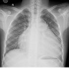
Trouble. Chest X-ray of the patient shows gross cardiomegaly.

Double
Trouble. CT angiography MPR image displays the thin hypodense band in the left ventricular outflow tract consistent with subaortic membrane. LV: Left ventricle.

Double
Trouble. CT angiography axial image displays the thin hypodense band in the left ventricular outflow tract consistent with subaortic membrane
subaortic membrane

