Tumoren des Fußes

MRI imaging
of soft tissue tumours of the foot and ankle. Angiomyoma. Axial T1W (a), sagittal T2FS (b) and post contrast T1FS (c) images of the ankle showing a well-defined lesion adjacent to the Achilles tendon which appears isointense on T1W, hyperintense on T2W and avidly enhances on postcontrast imaging. Note the capsule visible on T2FS (arrows)

MRI imaging
of soft tissue tumours of the foot and ankle. Lipoma. Axial T1W (a) and PDFS (b) images of the ankle demonstrating an encapsulated lesion (arrows). The lesion is isointense to fat on T1 with uniform loss of signal on PDFS consistent with a lipoma

MRI imaging
of soft tissue tumours of the foot and ankle. Soft tissue chondroma. Axial T1W (a) and T2FS (b) images of the ankle with a large well-defined posteromedial mass (arrows) which appears T1 hypointense and T2 hyperintense with internal low signal chondroid foci (arrowheads). The chondroid matrix is readily appreciated on the corresponding radiograph (c)

MRI imaging
of soft tissue tumours of the foot and ankle. Synovial sarcoma. Short axis T2FS image of the foot (a) showing a heterogeneous T2 hyperintense mass on the plantar aspect of the foot with heterogeneous enhancement of the viable tumour on the sagittal post-contrast T1FS image (b)

MRI imaging
of soft tissue tumours of the foot and ankle. Rhabdomyosarcoma. Short axis T1 image (a) of the foot of a 14-year-old girl demonstrating a hyperintense mass in the first web-space with avid enhancement on postcontrast T1FS imaging (b)

MRI imaging
of soft tissue tumours of the foot and ankle. Kaposi’s sarcoma. Coronal T1W (a) and post-contrast T1FS (b) images of the foot showing an exophytic lesion, low signal on T1W with avid enhancement and a nodular proliferation of the dermis

MRI imaging
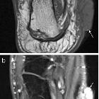
of soft tissue tumours of the foot and ankle. Extraosseous Ewing’s sarcoma. a Coronal T1W and (b) sagittal T2FS images of the foot. The lesion is isointense on T1W and hyperintense on T2FS and lies superficial to the plantar fascia

MRI imaging
of soft tissue tumours of the foot and ankle. Lymphoma. Axial T1FS (a) and post-contrast T1FS (b) images of the ankle demonstrating an infiltrative process surrounding the posterior tendons and neurovascular structures, isointense to skeletal muscle on T1W with mild diffuse contrast enhancement
 Assoziationen und Differentialdiagnosen zu Tumoren des Fußes:
