patellar tumors

Primary
tumors of the patella. Patellar giant cell tumor. (A, B) An enlargement of the cystic lesion in the patella and a bone translucency with peripheral rim change can be seen at the lateral down part of the patella on radiographs. (C, D) CT indicates that there is a well-defined lytic lesion with the thin cortex occupying two-thirds of the patella.

Primary
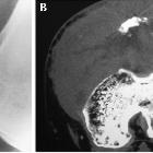
tumors of the patella. Chondroblastomas of the patella. (A) On plain radiographs, the lesion appears as geographic changes with lobulated margins, thinning cortex, sclerotic rim, and reactive bone. (B) Calcifications inside the lesion are seen on CT images.

Primary
tumors of the patella. An intracortical osteoid osteoma of the patella. (A) On plain radiographs, a clear lesion with small fuzzy periosteal reaction is observed. (B) CT demonstrates the sclerosis surrounding the lesion.

Primary
tumors of the patella. Patellar hemangioma. (A) Radiographs show a lytic lesion with sharp margins, thinning cortex, and sclerotic rim. (B, C) CT results accord with the radiographic results.

Primary
tumors of the patella. Osteosarcoma of the patella. (A) Plain radiograph and (B, C) CT images reveal a lytic lesion in the patella with partial cortical disruption.

Primary
tumors of the patella. Patellar lymphoma with a pathologic fracture. (A) On radiographs, the patella consists of a sclerotic proximal portion and a lytic distal portion. (B) Permeative bone destruction, hazy margins, destroyed cortex, large soft tissue mass surrounding the patella, and joint involvement are shown on CT images.

Primary
tumors of the patella. Patellar leiomyosarcoma. (A) Plain radiograph shows a mixed lytic and sclerotic lesion of patella. The margin of the lesion is ill defined and associated with cortical breach. (B) CT scan reveals multiple lytic lesions of the patella with a sclerotic rim and cortical disruption.

Primary
tumors of the patella. Hemangioendothelioma of the patella. Septation and thinning cortex in the osteolytic lesion are observed on (A) plain radiographs and (B) CT images.

Uncommon
cause for anterior knee pain - Aggressive aneurysmal bone cyst of the patella. Magnetic resonance imaging of the left knee. MRI of the left knee showing a hyperintense lesion within the inferior pole of the patella. (a) sagittal T1, (b) axial T1, and (c) axial T2 weighted sequences.

Uncommon
cause for anterior knee pain - Aggressive aneurysmal bone cyst of the patella. Computed tomography of the left knee. CT scan of the left patella showing an osteolytic lesion within the inferior pole of the patella. Note the cortical thinning/breakthrough as a sign for the aggressiveness of the lesion. (a) sagittal, (b) axial sequence.
Patellar tumors are extremely rare. They can be either benign or malignant primary bone tumors, or metastases.
Epidemiology
Patellar tumors represent just 0.1% of all primary bone tumors .
Clinical presentation
Patients may present with anterior knee pain and/or a palpable mass .
Pathology
Etiology
Most (~80%) patellar tumors are benign. A non-extensive list of causes is :
- benign
- giant cell tumor (most common)
- chondroblastoma (second most common)
- aneurysmal bone cyst
- malignant
Siehe auch:

 Assoziationen und Differentialdiagnosen zu Tumoren der Patella: