pulmonale Metastasen bei hepatozellulärem Karzinom
Teenager with
a liver mass. CXR AP shows multiple large round non-calcified lesions in the lungs bilaterally.The diagnosis was lung metastases in a patient with fibrolamellar hepatocellular carcinoma.

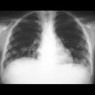
Pulmonary
metastases from primary hepatocellular carcinoma in a 26-year-old patient: a case report. Chest X-ray showing multiple variable sized nodules predominating in the inferior areas of the lungs.

Pulmonary
metastases from primary hepatocellular carcinoma in a 26-year-old patient: a case report. Computed tomography of the lungs, with pulmonary (A and B) and mediastinal (C and D) window settings, showing well-defined nodules, in a peripheral distribution.

Pulmonary
metastases from primary hepatocellular carcinoma in a 26-year-old patient: a case report. Computed tomography scans of the upper abdomen showing hepatomegaly, with an extensive ill-defined heterogeneous lesion in the hepatic parenchyma.

Artificial
pneumothorax improves radiofrequency ablation of pulmonary metastases of hepatocellular carcinoma close to mediastinum. Artificial pneumothorax adjuvant RFA of pulmonary metastases contiguous to the mediastinum (a 67-year-old man with a metastatic lesion in the superior lobe forepart of right lung). Tumor size, 1.5 × 1.2 cm. a Before ablation, chest CT imaging was performed to evaluate the anatomic relationship between tumor and peripheral cardiovascular structures. b Subsequently, a 22-G needle tip was used to create a puncture that reached the outer edge of pleura for injection of 1–2 ml saline. c The needle tip entered into the pleura, and the saline in the tube flowed into the cavity. d-e CO2 gas was administered gradually with a syringe until the tumor was separated from the mediastinum. f CT image during RFA showed the electrode inserted into the tumor and located away from the mediastinum by proxy of artificial pneumothorax. g The ablation zone gradually increased following the RFA procedure. h After RFA, the pulmonary texture around tumor showed a circular exudation shadow with ground-glass appearance on CT image. i Contrast enhanced CT image 1 month after RFA showed no enhancement of the ablated tumor contiguous to the mediastinum. j-l The size of ablated tumor decreased gradually after RFA during follow up at 3, 6, and 12 months, respectively
pulmonale Metastasen bei hepatozellulärem Karzinom
Siehe auch:


 Assoziationen und Differentialdiagnosen zu pulmonale Metastasen bei hepatozellulärem Karzinom:
