subdurale Verkalkung

Subdural
hemorrhage • Chronic calcified subdural hemorrhages - Ganzer Fall bei Radiopaedia

Subdural
hemorrhage • Chronic calcified subdural hemorrhages - Ganzer Fall bei Radiopaedia

Subdural
hemorrhage • Calcified chronic subdural hematoma - Ganzer Fall bei Radiopaedia

Symptomatic
calcified chronic subdural hematoma in an elderly patient: a case report. Pre- and post-contrast computerized tomography scan showing calcified subdural hematoma

Postoperative
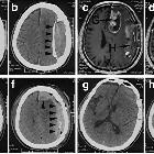
hemorrhage in an elderly patient with a glioblastoma multiform and a calcified chronic subdural hematoma. The preoperative and postoperative images of CT and MRI. (a,b) The CT images display the CSDH. The arrowheads point to the calcified membranes. (c, d) The MRI images show the GBM (G) and the CSDH (H). (e, f) The CT images indicate the in-situ hemorrhages after removal of the GBM and CSDH. (g, h) The postoperative CT images of the secondary craniotomy.

Calcified
chronic subdural hematoma • Calcified chronic subdural hematoma - Ganzer Fall bei Radiopaedia

Calcified
chronic subdural hematoma • Chronic overshunting - Ganzer Fall bei Radiopaedia

Calcified
chronic subdural hematoma • Calcified chronic subdural hematoma - Ganzer Fall bei Radiopaedia

 Assoziationen und Differentialdiagnosen zu subdurale Verkalkung: