Plantar xanthoma

Imaging of
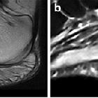
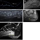
plantar fascia disorders: findings on plain radiography, ultrasound and magnetic resonance imaging. Plantar xanthoma. On both sagittal T1-weighted (a) and fluid-sensitive (b) images, xanthoma (arrows) appears as fusiform enlargement of the PF and shows heterogeneous signal intensity
Plantar xanthoma is a condition when xanthomatous deposits are occurring within the plantar fascia.
Clinical presentation
They are usually asymptomatic, although in some instances may result in vague pain and may also have unfavorable cosmetic effects.
Pathology
Xanthomas consist of localized collections of tissue histiocytes containing lipids, most frequently in the skin and subcutaneous tissues. As with xanthomas elsewhere, they are a feature of several types of primary hyperlipidemias.
Radiographic features
Plain radiograph
May be seen as soft-tissue masses without calcification.
MRI
Can show fusiform tendinous or aponeurotic enlargement with heterogeneous signal intensity
Can show
- small foci of increased tendinous signal intensity corresponding to xanthomatous deposits
- trabeculated low-signal-intensity areas representing the remaining tendinous fascicles
- these can produce a characteristic speckled or reticulated appearance on both T1- and T2images
Siehe auch:

 Assoziationen und Differentialdiagnosen zu Xanthome der Plantaraponeurose:

