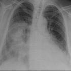
parenchymal lung disease
Parenchymal lung diseases can broadly be divided into those that create an abnormal increase in density on a chest radiograph and those that cause increased lucency.
The attenuation of any tissue on a radiograph is related to its density and in the lung, this is determined by the ratio of gas to surrounding soft tissue (blood, lung parenchyma or stroma) - normally 11 to 1. Any process that increases the amount of soft tissue creates a significant decrease in this ratio resulting in increased opacification. By virtue of the normal ratio of gas to soft tissue, this is more apparent on plain radiography than any process that decreases the amount of soft tissue, e.g. reduced blood flow, parenchymal or stromal destruction. CT, with its superior contrast resolution, is more sensitive when assessing overall decreases in radiographic density.
Pulmonary opacification
Abnormal pulmonary opacification may be subdivided into smaller groups by the pattern it creates on radiographic studies. These patterns have been shown to accurately represent the underlying pulmonary pathologic processes and are a practical way to generate a differential diagnosis. The patterns include:
- airspace opacification : consolidation
- atelectatic opacification : collapse
- interstitial opacification : lines
- nodular opacification : dots
Siehe auch:
- Konsolidierung der Lunge
- lung parenchyma
- nodular opacification
- pulmonary opacity
- interstitial opacification
und weiter:


 Assoziationen und Differentialdiagnosen zu