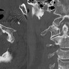
Cervical spine (odontoid view)

The odontoid or 'peg' projection is an AP projection of C1 (atlas) and C2 (axis).
Indications
This view focuses primarily on the odontoid process, and is useful in visualizing odontoid and Jefferson fractures.
Patient position
- patient positioned erect in AP position unless trauma the patient will be supine
- patient’s shoulders should be at equal distances from the image receptor to avoid rotation, the head facing straight forward.
- at the last instant, the patient is instructed to open their mouth as wide as possible
- the head should be positioned so the lower margin of the upper incisors and the base of the skull are perpendicular to the image receptor
- do not move the head in trauma, angle the central accordingly
Technical factors
- anterior-posterior projection
- centering point
- the central ray is centered at the center of the open mouth
- angle accordingly; see 'patient positioning'
- collimation
- laterally to include the mandible
- superior-inferior to include the upper incisors and lower incisors
- orientation
- landscape
- detector size
- 18 cm x 24 cm
- exposure
- 70-75 kVp
- 8-12 mAs
- SID
- 100 cm
- grid
- yes
Image technical evaluation
- the dens is free from superimposition of the adjacent atlas lateral masses or other tissues
- the zygapophyseal joint space between C1 and C2 is symmetrical
Practical points
- make sure that any removable artefacts such as earrings, glasses or metal dentures are removed to avoid obscuring the anatomy of interest
Positional errors
Teeth superimposing the dens
If teeth are superimposed over the upper aspect of the dens, the head needs to be hyperextended or in the case of trauma, the central ray should be angled cephalic.
Skull base superimposing the dens
If the base of skull is superimposed over the upper aspect of the dens, the head needs to be hyperflexed or in the case of trauma, the central ray should be angled caudally.
Siehe auch:

 Assoziationen und Differentialdiagnosen zu Cervical spine (odontoid view):


