Komplikationen Nabelarterienkatheter

Premature
newborn after umbilical arterial catheter placement. CXR shows the tip of the umbilical arterial catheter to be at the level of the L2 vertebral body.The diagnosis was umbilical arterial catheter malfunction with the tip of the catheter being low in position.

Premature
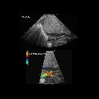
newborn now 2 weeks old with sepsis who had an umbilical arterial catheter placed at birth which was removed after a weekSagittal and transverse grayscale US of the aorta shows an echogenic, linear object within the aorta which on color doppler US was seen to have blood flow around it.The diagnosis was non-occlusive thrombus of the aorta secondary to past umbilical arterial catheterization.

Premature
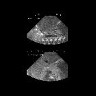
newborn now 1 month old with decreased urine output who had an umbilical arterial catheter placed at birthCoronal grayscale US of the aorta centered at the level of the kidneys (above) shows a round echogenic object within the aorta at the level of the origin of the renal arteries. Coronal color doppler US of the aorta (below) shows good blood flow around the object and into the renal arteries which was confirmed on spectral doppler US.The diagnosis was non-occlusive thrombus of the aorta secondary to past umbilical arterial catheterization.

Premature
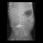
newborn after umbilical arterial catheter placement. AXR AP shows an umbilical arterial catheter whose tip projects inferiorly over the left external iliac artery. The tip of the umbilical venous catheter is at the cavo-atrial junction.The diagnosis was umbilical arterial catheter malfunction with malposition of the tip in the left external iliac artery.
Komplikationen Nabelarterienkatheter
Siehe auch:

 Assoziationen und Differentialdiagnosen zu Komplikationen Nabelarterienkatheter:
