okzipitale Dermoidzyste mit Dermalsinus

Cerebellar
abscess secondary to occipital dermoid cyst with dermal sinus. Sagittal T2, FLAIR images showing perilesional oedema in cerebellum and communication.

Cerebellar
abscess secondary to occipital dermoid cyst with dermal sinus. Sagittal View MRI Brain, T2, FLAIR images showing perilesional oedema in cerebellum and communication.

Cerebellar
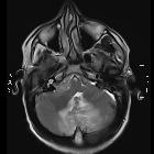
abscess secondary to occipital dermoid cyst with dermal sinus. Axial T2W image, lesion in midline in vermis region perilesional oedema and mass effect over 4th ventricle with hydrocephalus. Dermal sinus tract through occipital bone defect to scalp dermoid with secondary infection.

Cerebellar
abscess secondary to occipital dermoid cyst with dermal sinus. Axial T2W image, lesion in midline with perilesional oedema and mass effect. Communication/dermal sinus tract through occipital bone defect to scalp dermoid with secondary infection. Incidental detected right sided acoustic schwannoma.

Cerebellar
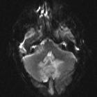
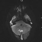
abscess secondary to occipital dermoid cyst with dermal sinus. MRI Brain, axial view, diffusion weighted image showed restricted diffusion.

Cerebellar
abscess secondary to occipital dermoid cyst with dermal sinus. MRI Brain, axial view, diffusion weighted image showed restricted diffusion.

Cerebellar
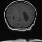
abscess secondary to occipital dermoid cyst with dermal sinus. Pre-contrast T1W coronal images – high signal in subarachnoid space in posterior fossa consistent with leakage of fatty content of dermoid.

Cerebellar
abscess secondary to occipital dermoid cyst with dermal sinus. Pre-contrast T1W coronal images – high signal in subarachnoid space in posterior fossa consistent with leakage of fatty content of dermoid.

Cerebellar
abscess secondary to occipital dermoid cyst with dermal sinus. Pre contrast T1W image shows multiple foci in posterior and midline region of cerebellar folia in keeping with leakage of fat content from high signal dermoid cyst in occipital region of scalp through sinus.

Cerebellar
abscess secondary to occipital dermoid cyst with dermal sinus. Pre contrast T1W image shows multiple foci in posterior and midline region of cerebellar folia in keeping with leakage of fat content from high signal dermoid cyst in occipital region of scalp through sinus.

Cerebellar
abscess secondary to occipital dermoid cyst with dermal sinus. Post-contrast T1W MR shows high signal foci which were non-enhancing suggesting leakage of fat containing dermoid cyst in cerebellar folia.

Cerebellar
abscess secondary to occipital dermoid cyst with dermal sinus. Post-contrast coronal T1W images showed ring enhancement around focal collection.

Cerebellar
abscess secondary to occipital dermoid cyst with dermal sinus. Post-contrast coronal T1W images showed ring enhancement around focal collection.
okzipitale Dermoidzyste mit Dermalsinus
Siehe auch:

 Assoziationen und Differentialdiagnosen zu okzipitale Dermoidzyste mit Dermalsinus:

