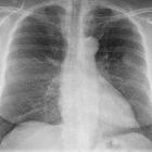
Lipom der Thoraxwand


Chest wall lipomas are benign fat containing thoracic lesion.
Epidemiology
While they can occur at any age, they typically occur in older patients who are 50-70 years of age, and they are most frequent in those with increased an increased body mass index.
Pathology
They are well-circumscribed encapsulated masses composed of adipocytes that differ very little from normal fatty tissue. Most lipomas that originate in the chest wall are deep lipomas, which tend to be larger and less well circumscribed than superficial lesions
Radiographic features
CT
Follows homogenous fat attenuation in general. However, multiple thin septa often are present that appear slightly enhanced on CT scans.
MRI
Lipomas generally appear to be internally homogeneous and do not enhance after intravenous contrast material administration. They typically follows fat signal on all sequences.
- T1: can have high signal
- T1 fat sat: shows fat suppression and has low signal intensity
Siehe auch:

 Assoziationen und Differentialdiagnosen zu Lipom der Thoraxwand:


