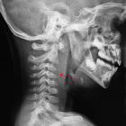
retropharyngealer Desmoidtumor

A rare case
of desmoid tumour within the retropharyngeal and prevertebral spaces. Sagittal CT showing a mass within the retropharyngeal and prevertebral spaces, isodense compared to the muscle (a), and enhancing after contrast administration (b), with bone erosion (c).

A rare case
of desmoid tumour within the retropharyngeal and prevertebral spaces. Axial CT: the mass is inseparable from prevertebral musculature, displaces and compresses the trachea and deviates the carotid artery laterally.

A rare case
of desmoid tumour within the retropharyngeal and prevertebral spaces. Coronal T1 weighted shows an isointense tumour compared to muscle (a), hypointense on T2W (b), with avid post-contrast enhancement (c).
retropharyngealer Desmoidtumor
Siehe auch:

 Assoziationen und Differentialdiagnosen zu retropharyngealer Desmoidtumor:
Desmoid-Tumor
- aggressive Fibromatose


