Impingement Syndrome bei Patienten mit multiplen kartilaginären Exostosen

Characteristics
of hip impingement syndrome in patients with multiple hereditary exostoses. Measurement of the radiographic alpha angle. The alpha angle is determined by the angle between a line from the center of the femoral head through the middle of the femoral neck and a line through a point where the contour of the femoral head-neck junction exceeds the radius of the femoral head

Characteristics
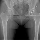
of hip impingement syndrome in patients with multiple hereditary exostoses. Plain radiographic measurements to evaluate the hip joint deformities. a The femoral neck-shaft angle is determined by measuring the angle created by a line in the central axis of femoral shaft and a second line created by the connection of the femoral head center to the mid portion of the femoral head and neck junction contour. b Sharp’s angle is determined by measuring the angle created by a line connecting the acetabular tear drops and a second line connecting a tear drop and the sourcil end. c The center-edge angle is determined by measuring the angle created by a line connecting the vertical line to the tear drop line through the center of the femoral head and a second line from the center of the hip to the lateral acetabular wall margin

Characteristics
of hip impingement syndrome in patients with multiple hereditary exostoses. Measurement of minimum ischio-femoral distance to evaluate ischio-femoral impingement in a computed-tomographic study. The minimum ischio-femoral distance is determined by the nearest distance between the exostoses and ischium around the lesser trochanter area were measured at axial CT plane

Characteristics
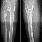
of hip impingement syndrome in patients with multiple hereditary exostoses. Illustration of the hip joint explaining why ischio-femoral impingement (IFI) symptoms are more common than femoro-acetabular (FAI) symptoms in hips with multiple hereditary (MHE). a The normal hip joint morphology without MHE. The hatched part in the figure shows the exostoses that can occur in the femoral head and neck area. In this circumstance, the incidence of both FAI and IFI is likely to increase. b MHE hip patients with the characteristic deformities. Though hatched part of the exostoses may increase the possibility of the impingement, coxa valga deformity reduces the possibility of impingement between the femoral head and acetabulum by increasing the working distance. On the contrary, coxa valga deformity acts as a risk factor for the development of IFI reducing the distance between the exostoses and the ischium
Impingement Syndrome bei Patienten mit multiplen kartilaginären Exostosen
Siehe auch:
- Multiple kartilaginäre Exostosen
- kartilaginäre Exostose Femur
- ischiofemorales Impingement
- kartilaginäre Exostosen Becken
und weiter:

 Assoziationen und Differentialdiagnosen zu Impingement Syndrome bei Patienten mit multiplen kartilaginären Exostosen:


