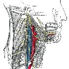
Hypoglossusparese

Hypoglossusparese
links als Folge einer osteoblastischen Metastasierung um das Foramen nervi hypoglossi bei Prostatakarzinom.

Persistent
idiopathic unilateral isolated hypoglossal nerve palsy – a report of two cases. a Right-sided atrophy along with tongue deviation 7 months after onset of symptoms. b Cisternal segment of the right hypoglossal nerve (HN) on 3D constructive interference in steady state (CISS) magnetic resonance imaging (MRI) showing normal appearance (white arrow). c Normal morphology of the skull base segment of the right HN inside of the right hypoglossal canal (HC) on 3D T1-weighted contrast enhanced (CE) MRI. The HN is of hypointense signal (white arrow), while the surrounding venous plexus shows prominent CE. d Axial computed tomography scan of the skull base demonstrating a normally appearing HC (white arrow). e Head and neck T1-weighted MRI demonstrates fatty infiltration and atrophy of the right half of the tongue due to chronic muscle denervation (white arrow)

Unilateral
tongue atrophy due to paralysis of the left hypoglossal nerve. Right tongue-muscle is normal (X). Computed Tomography.


 Assoziationen und Differentialdiagnosen zu Hypoglossusparese: