abgehängter isolierter Pankreasgang nach nekrotisierender Pankreatitis

Disconnected
pancreatic duct syndrome: imaging findings and endoscopic treatment. MRCP showed mild dilatation of the Wirsung duct at the pancreatic head (arrow), and segmental discontinuity (thick arrow) of the main pancreatic duct in the body. Normal appearance of the biliary tract.

Disconnected
pancreatic duct syndrome: imaging findings and endoscopic treatment. MRCP showed mild dilatation of the Wirsung duct at the pancreatic head (arrow), and segmental discontinuity (thick arrow) of the main pancreatic duct in the body. Normal appearance of the biliary tract.

Disconnected
pancreatic duct syndrome: imaging findings and endoscopic treatment. Repeated ERCP confirmed MRCP findings of mild dilatation of the Wirsung duct in the pancreatic head (arrow), and focal discontinuity (thick arrow) of the main pancreatic duct in the body.

Disconnected
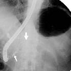
pancreatic duct syndrome: imaging findings and endoscopic treatment. Definitive endoscopic treatment required positioning of a long pancreatic stent (arrowhead) through the main pancreatic duct discontinuity. Note mild dilatation of the Wirsung duct in the pancreatic head (arrow),

Disconnected
pancreatic duct syndrome: imaging findings and endoscopic treatment. Cysto-gastrostomy with metallic stent (thin arrows) and nasal-cystic tube (arrows) was previously positioned to drain the major peripancreatic collection (*).

Disconnected
pancreatic duct syndrome: imaging findings and endoscopic treatment. Cysto-gastrostomy with metallic stent (thin arrows) and nasal-cystic tube (arrows) was previously positioned to drain the major peripancreatic collection (*).

Disconnected
pancreatic duct syndrome: imaging findings and endoscopic treatment. Maximum-intensity projection (MIP) images showed the metallic stent (thin arrows) and nasal-cystic tube (arrows) previously positioned at the cysto-gastrostomy.

Disconnected
pancreatic duct syndrome: imaging findings and endoscopic treatment. Unenhanced (a) and post-contrast (b,c) images showed reappearance of the major post-necrotic fluid collection (*) measuring approximately 9x2.5 cm, located above the pancreatic body-tail (+).

Disconnected
pancreatic duct syndrome: imaging findings and endoscopic treatment. Unenhanced (a) and post-contrast (b,c) images showed reappearance of the major post-necrotic fluid collection (*) measuring approximately 9x2.5 cm, located above the pancreatic body-tail (+).

Disconnected
pancreatic duct syndrome: imaging findings and endoscopic treatment. The pancreatic body and tail (+) were recognizable with preserved anatomic continuity and enhancement. The main pancreatic duct was not perceptible.

Disconnected
pancreatic duct syndrome: imaging findings and endoscopic treatment. Axial T1-(a), T2-(b), diffusion-weighted (c) and apparent diffusion coefficient (ADC) map (d) showed stable major post-necrotic fluid collection (*) measuring approximately 9x2.5 cm, located above the pancreatic body-tail.
abgehängter isolierter Pankreasgang nach nekrotisierender Pankreatitis
Siehe auch:

 Assoziationen und Differentialdiagnosen zu abgehängter isolierter Pankreasgang nach nekrotisierender Pankreatitis: