abgerissener ZVK in Pulmonalarterie
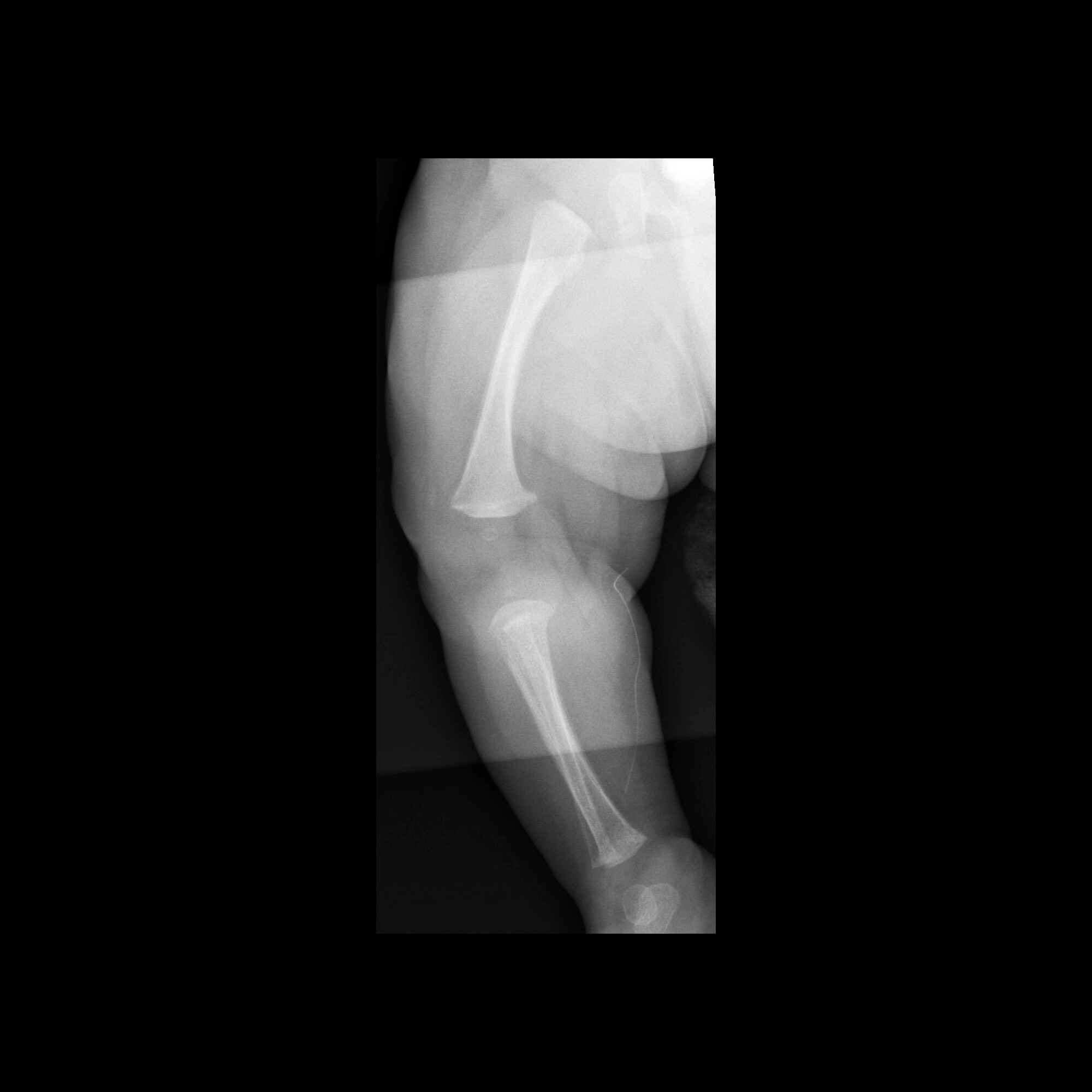
Infant after
left lower extremity PICC removal which after measuring was found to be too short in length. AXR AP (left) and lateral (right) show a discontinuous catheter fragment in the left femoral and iliac veins.The diagnosis was peripherally inserted central venous catheter malfunction with the catheter having broken during removal with a retained catheter fragment in the left femoral and iliac veins.
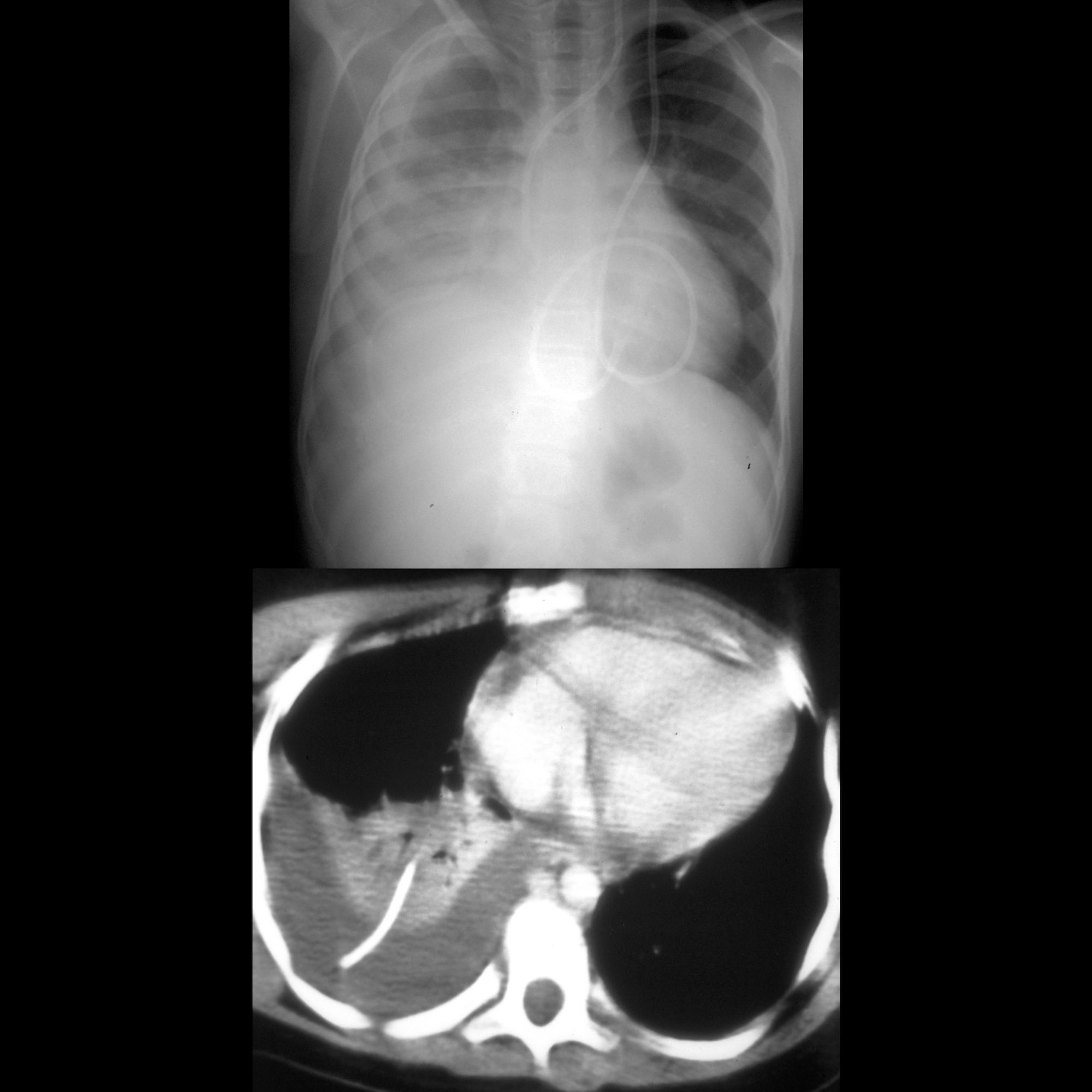
School ager
who presents with back pain after having a central venous catheter replaced 5 months agoCXR AP shows the tip of the new catheter to be in appropriate location in the superior vena cava. There is a catheter fragment in the right lower lobe along with a large right pleural effusion. Axial CT with contrast of the chest shows the embolized catheter fragment in the periphery of the right lower lobe and to have eroded into the right pleural space, with an associated pleural effusion and atelectasis.The diagnosis was an embolized catheter fragment after catheter removal.
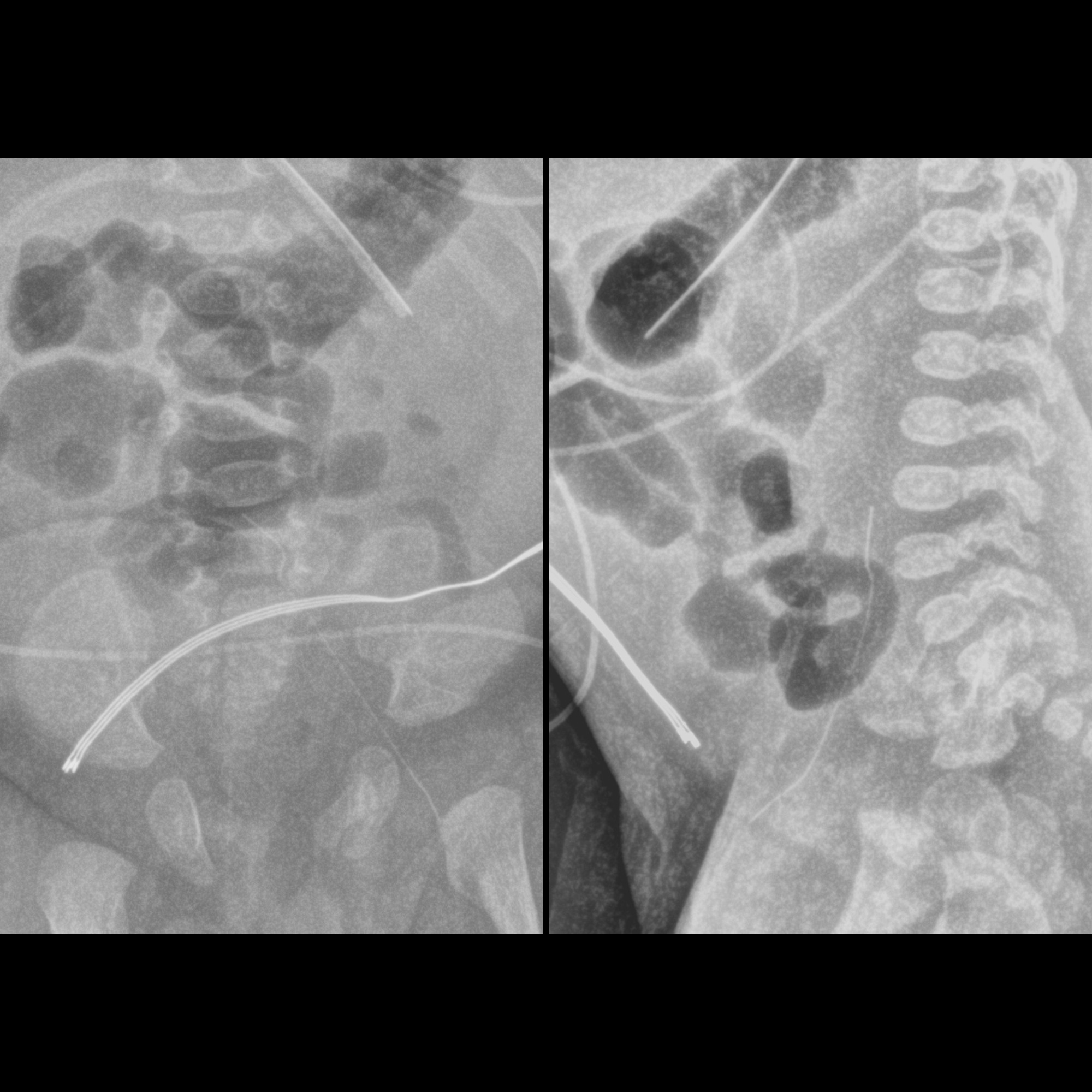
Infant who
has just had a right lower extremity PICC line removed which upon measuring was seen to be shorter than it should have been. AP radiograph of the right lower extremity shows a retained fragment of a PICC line in the posterior soft tissues of the right calf.The diagnosis was peripherally inserted catheter malfunction with a retained fragment of the removed PICC line in the right calf.


 Assoziationen und Differentialdiagnosen zu abgerissener ZVK:

