Adenocarcinoma of the lacrimal glands


Adenocarcinoma of the lacrimal glands is rare, with few cases reported in the literature since it was first described in 1996 . Primary adenocarcinoma of the lacrimal gland is extremely rare; only 9 cases have been reported in the literature . It can be classified into high- and low-grade malignancies. This neoplasm is histologically and immunohistochemically similar to salivary ductal carcinoma .
Clinical presentation
They usually present as unilateral gland enlargement with displacement of the eye.
Pathology
Histologic examination shows a well-circumscribed but unencapsulated neoplasm. The cells are highlighted by the calponin immunostain, consistent with myoepithelial differentiation. These cells have round to oval nuclei with a uniform vesicular chromatin pattern and small nucleoli. Most of these cells exhibit cytoplasmic and membranous staining for C-kit (CD117). p53 expression is seen in a minority of the nuclei. A significant number of luminal structures contain mucicarmine-positive material. The tumor cells are uniformly negative for GFAP.
Radiographic features
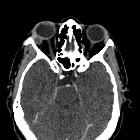
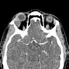
A rounded extraconal mass centered around the lacrimal gland in the superolateral aspect of the orbit.
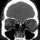
CT
Axial and coronal CT shows peripheral and mild central heterogeneous enhancement.
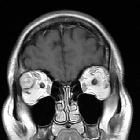
MRI
Signal characteristics include:
- T1: intermediate intensity lacrimal fossa mass
- T2: usually hyperintense
- T1 C+: heterogeneous enhancement
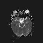
- DWI and ADC: diffusion restriction of the peripherally enhancing component
Treatment and prognosis
The tumor is usually treated with complete excision, lymph node dissection, and adjuvant radiotherapy. Because there are few reports of this entity, its clinical behavior, prognosis and definitive treatment are still unknown .
Differential diagnosis
Possible differential considerations include:
- lacrimal gland sarcoidosis
- orbital pseudotumor
- lacrimal gland pleomorphic adenoma
- lacrimal gland oncocytoma
- lacrimal gland adenoid cystic carcinoma
- lacrimal gland malignant lymphoma
- benign reactive lymphoid hyperplasia
See also
Siehe auch:


 Assoziationen und Differentialdiagnosen zu Adenokarzinom der Glandula lacrimalis:


