Aneurysma der Vena subclavia

Subclavian
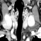
vein aneurysm - Case presentation and discussion. Neck and thoracic inlet CT with intravenous contrast media in axial projection, a saccular aneurysm of the upper edge of the left subclavian vein can be seen (arrow), without evidence of rupture.
Subclavian
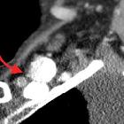
vein aneurysm - Case presentation and discussion. Neck and thoracic inlet CT with intravenous contrast media in coronal reconstruction, in which a saccular aneurysm of the upper edge of the left subclavian vein can be seen (arrow), without evidence of rupture.
Subclavian
vein aneurysm - Case presentation and discussion. Neck and thoracic inlet CT with intravenous contrast media in sagittal reconstruction, in which a saccular aneurysm of the upper edge of the left subclavian vein can be seen (arrow), without evidence of rupture.
Subclavian
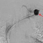
vein aneurysm - Case presentation and discussion. Venography of the left subclavian vein after catheterization and subsequently of the aneurysm, shows filling of the venous dilatation after the injection of contrast media, with preserved patency of the subclavian vein.
Subclavian
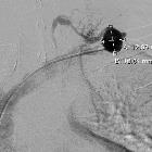
vein aneurysm - Case presentation and discussion. Venography of the left subclavian vein after catheterization and subsequently of the aneurysm, shows filling of the venous dilatation after the injection of contrast media. The dimensions of the lesion are noted.
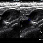
Subclavian
vein aneurysm - Case presentation and discussion. Gray scale and Color Doppler exploration demonstrates the aneurysm and an echogenic image attached to a wall in its interior, compatible with mural thrombus.

Subclavian
vein aneurysm - Case presentation and discussion. Gray scale and Color Doppler exploration, orthogonal to Fig. 3a, demonstrates the aneurysm and an echogenic image attached to a wall in its interior, compatible with mural thrombus.
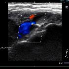
Subclavian
vein aneurysm - Case presentation and discussion. Gray scale and Color Doppler exploration, showing evidence of the patency of the subclavian vein adjacent to the aneurysm.
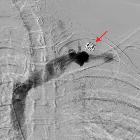
Subclavian
vein aneurysm - Case presentation and discussion. Venography of the left subclavian vein after endovascular therapy with coils within the aneurysm. Complete exclusion of aneurysm from the venous circulation and patency of the veins is observed.
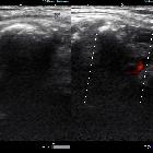
Subclavian
vein aneurysm - Case presentation and discussion. Power Doppler demonstrates irregular echogenic image which generates acoustic shadowing, corresponding to embolisation material within the aneurysm. There is adequate bloodflow within the subclavian vein.
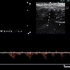
Subclavian
vein aneurysm - Case presentation and discussion. Spectral Doppler demonstrates adequate bloodflow within the subclavian vein.
Aneurysma der Vena subclavia
Siehe auch:

 Assoziationen und Differentialdiagnosen zu Aneurysma der Vena subclavia:

