anorektale Malformation

Newborn with
a defect of the lower abdominal wallAXR AP shows diastasis of the symphysis pubis and multiple spinal segmentation defects while the AXR lateral shows a small amount of bowel herniated anterior to the abdomen and inferior to the umbilicus along with a large skin covered spinal dysraphism posteriorly and a radioopaque marker being held in place over where the anus should be.The diagnosis was cloacal exstrophy with bladder exstrophy, anorectal malformation and spinal segmentation anomalies.

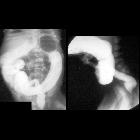
Newborn with
failure to pass meconium. AXR (above) shows multiple dilated loops of bowel within the abdomen that are progressively more dilated as they near the rectum. Prone cross-table lateral radiograph of the abdomen obtained after 10 minutes in the prone position (below) with a radio-opaque BB on the anus shows a short distance between the anus and the gas in the rectum.The diagnosis was a low anorectal malformation.

Newborn with
an absent anal orifice. Surgical image shows the rectum opened posteriorly through a posterior sagittal incision. A small catheter (in the center) is shown coursing through a rectourethral fistula.The diagnosis was anorectal malformation.

Newborn with
absent anus and stool coming out of the vagina. CXR AP (left) shows a hemivertebra at L1 causing spinal curvature convex left. Transverse US of the pelvis (above right) shows in the midline anteriorly an anechoic fluid-filled bladder with a round echogenic stool-filled rectum posterior to it while a transverse US of the perineum (below right) shows a very short distance between the calipers superiorly on the skin and inferiorly on the anterior wall of the rectum. The diagnosis was low anorectal malformation and congenital scoliosis.


 Assoziationen und Differentialdiagnosen zu anorektale Malformation: