Anterior abdominal wall

The anterior abdominal wall forms the anterior limit of the abdominal viscera and is defined superiorly by the xiphoid process of the sternum and costal cartilages and inferiorly by the iliac crest and pubic bones of the pelvis.
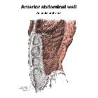
Gross anatomy
In general, the anterior abdominal wall has nine layers (from superficial to deep):
Scarpa's fascia is deep to the skin and subcutaneous fat in the lower part of the wall and is fused with Colle's fascia in the perineum.
The muscle layers include the external oblique muscle, internal oblique muscle, transversus abdominis muscle anterolaterally and the rectus abdominis muscle anteriorly. The fascia surrounding the 3 anterolateral muscles fuse anteriorly to attach to the rectus abdominis at the linea semilunaris. The fascia then continues medially surrounding the rectus abdominis as the rectus sheath further fusing in the midline with the contralateral fascia at the linea alba. Thus the combination of muscle, aponeuroses, and fascial layers form a corset-like structure that protects the abdominal viscera.
The arcuate line demarcates an important boundary on the anterior abdominal wall as the inferior limit of the rectus sheath. Superior to the arcuate line, the internal oblique aponeurosis surrounds the rectus abdominis muscle on its anterior and posterior aspects. Inferior to the arcuate line, the aponeuroses of internal oblique and transversus abdominis fuse and pass anteriorly to the rectus abdominis muscle.
Arterial supply
The muscles and associated soft tissues derive blood supply from branches of the superior epigastric, subcostal and inferior epigastric arteries and their cutaneous branches above the umbilicus.
Arterial supply below the umbilicus is from superficial epigastric arteries, superficial circumflex iliac arteries and superficial external pudendal arteries.
Venous drainage
Superficial veins are paired with the arteries. Veins above the umbilicus drain into the azygos system and below the umbilicus into the femoral system via the great saphenous vein.
Lymphatic drainage
The lymphatic vessels above the umbilicus drain into axillary and sternal nodes. The vessels below the umbilicus drain into superficial inguinal nodes.
Innervation
Derived from the ventral rami of T7 through L1. Thoracoabdominal nerves from the ventral rami of T7 to T11. Subcostal nerves from the ventral rami of T12. Ventral rami of the L1 nerve roots give rise to iliohypogastric and ilioinguinal nerves.
Variants
- Variable level of the arcuate line
- Variable number of horizontal septations in rectus abdominis
- Presence (or absence) of pyramidalis and/or rectus sternalis muscles
- Presence (or absence) of superficial inferior epigastric artery supplying superficial tissues of abdominal wall below the level of the umbilicus
Radiographic features
Plain radiograph
Muscle layers of the anterior abdominal wall may be outlined between the extraperitoneal fat and subcutaneous fat layers, especially in obese patients.
CT
Three muscle layers (external oblique, internal oblique, transverse abdominis) can be seen anterolaterally in cross section and also the rectus muscle and its sheath can be seen anterior to the other three muscle layers.
Related pathology

