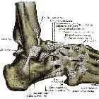
Anterior talofibular ligament injury
Anterior talofibular ligament injury is the most common of the ligament injuries that can occur as part of the lateral ligament complex injuries . The injuries can comprise either soft tissue tears, avulsion fractures or both.
Pathology
Anterior talofibular ligament injuries typically occur with an inversion injury to the ankle, either with or without plantar flexion. Approximately two-thirds of ankle sprains tend to be isolated injuries to the anterior talofibular ligament (ATFL), the weakest ligament in the lateral collateral complex of the ankle. There is general agreement that avulsion is more common at the fibular than the talar end of the ligament .
Grading
AMA classification :
- grade I: ligament stretch
- grade II
- partial tear
- partial loss of movement
- mild to moderate joint instability
- grade III
- complete tear
- inability to bear weight
- significant joint instability
Many orthopedists favor a functional approach based on clinical examination:
- grade I: stable injury
- provocative maneuvers do not elicit increased upper ankle joint laxity
- no ligament in the lateral complex is completely ruptured
- grade II: unstable
- ruptured anterior talofibular ligament
- increased upper ankle joint laxity
- grade III: unstable
- ruptured calcaneofibular ligament (CFL)
- increased upper ankle joint laxity
There are other grading systems, of course, such as the anatomic classification or grading by clinical presentation symptoms .
Radiographic features
Plain radiograph/CT
Many show an associated avulsion fracture at either the fibular end or, less commonly, the talar end.
May show ancillary features such as an ankle joint effusion and/or overlying lateral malleolar soft tissue swelling.
Ultrasound
Has been shown to have high sensitivity and specificity rates for chronic anterior talofibular ligament tears .
MRI
Both the anterior and posterior talofibular ligaments are usually seen on a single axial image obtained slightly distal to the tibiofibular ligaments .
MRI may show detachment, discontinuity, thickening, thinning, contour irregularity of the ligament, a bright rim sign or an associated bony avulsion. Heterogeneity with increased intraligamentous signal intensity on fat-suppressed T2-weighted or intermediate-weighted images is indicative of intrasubstance edema or hemorrhage.
Signal characteristics
- T2
- fluid within the joint serves to highlight the anterior talofibular ligament on T2-weighted images
- the ligament appears as a thin, straight, low signal intensity band extending from the talus to the fibular malleolus
- a bright rim sign (cortical defect with a bright dot-like or curvilinear high signal intensity lesion) may be seen on T2 weighted imaging
Differential diagnosis
On MRI consider:
- anterolateral ankle impingement
- repetitive synovial inflammation secondary to chronic lateral ankle instability produces a soft-tissue “mass” consisting of hypertrophic synovial tissue and fibrosis within the lateral gutter
Siehe auch:
- Os subfibulare
- Bänder des Sprunggelenkes
- posterior talofibular ligament
- Ligamentum fibulotalare anterius
- Ligamentum fibulocalcaneare
- Bandverletzungen am Außenknöchel
- Ligamentum tibiofibulare posterius
- lateral collateral ligament of the ankle
- Ruptur Ligamentum fibulotalare posterius
- Ruptur Ligamentum fibulocalcaneare

 Assoziationen und Differentialdiagnosen zu Verletzungen Ligamentum fibulotalare anterius:


