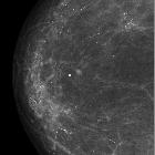
benigne neonatale Brustvergrößerung

Ultrasound of
neonatal breast enlargement. Ultrasound examination of the breast of the newborn: Shows retroareolar oval-shaped heterogenous masses (white arrows) that appeared of mixed echogenicity demonstrating hyperechoic central portion with multiple peripherally arranged variable-sized small cysts (yellow arrows).

Ultrasound of
neonatal breast enlargement. Follow-up ultrasound after two months shows remarkable regression of the size of the retroareolar tissues (white arrows) that appeared as a hypoechoic mass with asymmetric appearance, larger on the left side.

Ultrasound of
neonatal breast enlargement. Follow-up ultrasound examination of the breast after 6 months from the initial scan shows that there is a small remnant tissue in the retroareolar region resembling breast bud (white arrows).

 Assoziationen und Differentialdiagnosen zu benigne neonatale Brustvergrößerung:
