blutendes Jejunaldivertikel

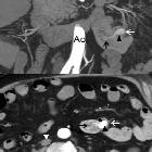
Massive lower
gastrointestinal bleeding from jejunal diverticulum with an occult Dieulafoy’s lesion. Coronal (upper) and axial (lower) images. Diverticulum (white arrow) originating in the jejunum wall (*), with stranding of the adjacent fat (white arrowhead). There was no hyperattenuating material within the diverticulum lumen.

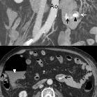
Massive lower
gastrointestinal bleeding from jejunal diverticulum with an occult Dieulafoy’s lesion. Coronal (upper) and axial (lower) images. Swirling jet of contrast material (black arrow) within the lumen of the diverticulum (white arrow). A collapsed inferior cava vein indicates hypovolaemia (white arrowhead).

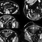
Massive lower
gastrointestinal bleeding from jejunal diverticulum with an occult Dieulafoy’s lesion. Coronal (upper) and axial (lower) images. The amount of intraluminal contrast increases in diverticular (black arrowhead) and jejunal (black arrow) lumen in comparision with arterial phase. Hyperattenuating material in the colon suggests blood (white arrowhead).

 Assoziationen und Differentialdiagnosen zu blutendes Jejunaldivertikel:
