bronchiale Tumoren

A rare cause
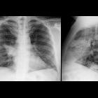
of post-obstructive pneumonia: endobronchial fibrolipoma. Chest X-ray (frontal and lateral projections) shows a right hilar opacity with indistinct borders, diagnosed as pneumonia of the right upper lobe.

A rare cause
of post-obstructive pneumonia: endobronchial fibrolipoma. Chest CT [axial (a and b) and sagittal sections (d)] shows post-obstructive pneumonia caused by a complete occlusion of the anterior segmental bronchus of the right upper lobe due to hypodense mass.

A rare cause
of post-obstructive pneumonia: endobronchial fibrolipoma. Chest CT [axial (a and b) and sagittal sections (d)] shows post-obstructive pneumonia caused by a complete occlusion of the anterior segmental bronchus of the right upper lobe due to hypodense mass.

Endobronchial
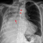
angiofibroma in the aberrant tracheal bronchus presenting as spontaneous pneumomediastinum. a Initial chest X ray shows pneumomediastinum. b Bronchoscopy finding of a protruding and glistering tumour originating from the right side of the trachea. c Computed tomography showing an elongated endobronchial tumour in the accessory tracheal bronchus originating from the right side of the lower tracheal wall (black arrow, axial view). d Computed tomography showing a tumour located in the right upper lobe from the accessory tracheal bronchus (white arrow, coronal view)

Giant
endobronchial hamartoma resected by fiberoptic bronchoscopy electrosurgical snaring. CT scan demonstrating a vegetating neoplasm of the left main bronchus (white arrow) without signs of extrabronchial infiltration.
bronchiale Tumoren
Siehe auch:
und weiter:

 Assoziationen und Differentialdiagnosen zu bronchiale Tumoren:


