chronic subdural hematoma

Infant with
acute and chronic emesisAxial CT without contrast of the brain (upper left) shows bilateral large low density extra-axial fluid collections and a left frontal small high density extra-axial fluid collection. There is also prominence of the cortical sulci and the ventricular system. Axial T1 (upper right), T2 (lower left) and FLAIR (lower right) MRI without contrast of the brain better demonstrates the extra-axial fluid collections with the bilateral large collections being bright on T1 and T2 and the small left frontal collection being iso on T1 and dark on T2.The diagnosis was bilateral large chronic subdural hematomas with associated cerebral atrophy and a left frontal acute subdural hematoma in a child abuse patient.
Infant with
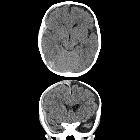
decreased responsivenessAxial (above) and coronal (below) CT without contrast of the brain shows a right moderate sized high density cresenteric extra-axial fluid collection that extends around the entire right cerebral hemisphere as well as interhemispherically. There is also a left moderate sized low-density cresenteric extra-axial fluid collection.The diagnosis was a right acute subdural hematoma and a left chronic subdural hematoma in a child abuse patient.

Ventriculoperitoneal
shunt complications: a local study at Qena University Hospital: a retrospective study. a Plain CT brain of an 18-month-old infant showing bilateral chronic subdural hematoma due to right-sided VP shunt overdrainage. b Plain CT brain of a 1-year-old infant showing right-sided chronic subdural hematoma due to right-sided VP shunt overdrainage


 Assoziationen und Differentialdiagnosen zu chronisches Subduralhämatom:

