congenital pulmonary lymphangiectasia

Newborn with
respiratory distress. CXR AP and lateral shows mild cardiomegaly and diffuse interstitial infiltrates with no evidence of pleural effusions.The diagnosis was pulmonary lymphangiectasia.

Congenital
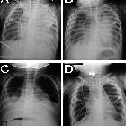
pulmonary lymphangiectasia. Pulmonary Lymphangiectasia. Chest radiographs, AP views. Radiological findings occurring during the clinical course of PL. A and B: over time progression of hazy perihilar infiltrates on the left lung. C: important bilateral pleural effusion. D: after pleurodesis, bilateral lung hyperinflation with interstitial and septa thickening are evident, and a mild degree of pleural effusion remains.

Congenital
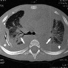
pulmonary lymphangiectasia. Pulmonary Lymphangiectasia. High-resolution computed tomography (HRCT). Diffuse thickening of the peribronchovascular interstitium and the interlobular septa (arrowheads), associated with bilateral pleural effusion (*), and peribronchovascular infiltrates (arrows) with bronchogram.

Congenital
pulmonary lymphangiectasia. Lymphoscintigraphy. Lymphoscintigraphic study of a patient affected by pulmonary lymphangiectasia and generalized lymphedema showed signs of dermal back-flow in the right lower limb. A and B = patient"s hands. L = liver. Arrows point to the thoracic duct.
Congenital pulmonary lymphangiectasia refers to congenital dilatation of the lymphatic system.
It typically presents in infancy. It may be isolated to lung or may be part of a generalized lymphangiectasia.
Siehe auch:
und weiter:


 Assoziationen und Differentialdiagnosen zu