COVID-19 myelitis
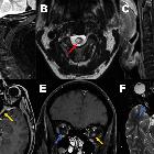
Myelin
oligodendrocyte glycoprotein antibody-associated optic neuritis and myelitis in COVID-19: a case report and a review of the literature. MRI sagittal STIR images (A) of the cervical spine reveal ill-defined long segment hyperintensity with prominent cord expansion C2–C4 (red arrow); B shows axial cut with cord signal alteration at C3 (red arrow), and C post-contrast sagittal image showed no abnormal enhancement. MRI orbit Axial T1-weighted fat suppression post-contrast (D) and coronal images (E) reveals abnormal enhancement of left optic nerve pre-chiasmatic (intracanicular); (yellow arrow) with corresponding hyperintense on axial T2 weighted images (F) (yellow arrow) with no abnormality of the right optic nerve (blue arrow)


 Assoziationen und Differentialdiagnosen zu COVID-19 myelitis: