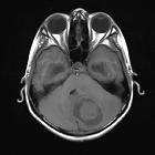
cystic cerebellar mass

Role of
diffusion-weighted imaging in differentiation between posterior fossa brain tumors. Epidermoid cyst in 38-year-old male complaining from auditory problems. A lobulated extra-axial cystic lesion is seen in the right cerebellopontine angle (CPA) insinuating itself, compressing the pons, extending into prepontine cistern, and encasing the basilar artery. It displays low T1WI signal intensity (a) and high T2WI signal intensity cerebrospinal fluid (CSF) like (b). On FLAIR, it shows low SI (partially suppressed SI (c). It shows faint marginal enhancement on post-contrast study (d). It shows iso SI-like brain parenchyma (restricted diffusion); it does not have high CSF SI-like arachnoid cyst (e)

School ager
with double vision. Axial CT with contrast of the brain shows a large heterogeneous cystic and solid mass encompassing the entire right cerebellar hemisphere which is compressing the slit-like fourth ventricle and shifting it to the left, resulting in obstructive hydrocephalus of the third and lateral ventricles.The diagnosis was cerebellar astrocytoma.

Cerebellar
pilocytic astrocytoma: MR spectroscopy. T2WI shows hyperintense lesion in posterior fossa with effaced 4th ventricle and triventricular hydrocephalus with eccenteric intermediate signal intensity solid component.

Magnetic
resonance imaging of pineal region tumours. Arachnoid cyst in a 27-year-old woman with headache and dizziness. Sagittal T2-weighted image (a) demonstrates a cystic infratentorial lesion (arrow) in the pineal recess, which has high signal. On coronal FLAIR image (b), the lesion (arrow) has isointense signal relative to CSF
zystische zerebelläre Raumforderungen
cystic cerebellar mass
Siehe auch:
- Mega Cisterna magna
- Arachnoidalzysten der hinteren Schädelgrube
- Kleinhirntumoren
- Hämangioblastom
- zystische Raumforderung hintere Schädelgrube
- zystische Malformationen der hinteren Schädelgrube
- zystische Tumoren im Kleinhirnbrückenwinkel
- zystische Läsionen des Gehirns
- zerebelläre Zystizerkose
- zystisches Schwannom des Kleinhirnbrückenwinkels
- atrial diverticula
- zystisches Meningeom im Kleinhirnbrückenwinkel
- Pilozytisches Astrozytom des Kleinhirns
- erweiterter retrocerebellärer Liquorraum
und weiter:

 Assoziationen und Differentialdiagnosen zu zystische zerebelläre Raumforderungen:
erweiterter
retrocerebellärer Liquorraum


