Dental implant

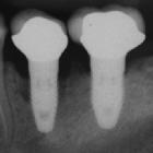
CT Scan for
Dental Implants. This shows eight dental implants superimposed over the lower jaw in areas of maximum bone and four teeth that will be extracted.
xray 2 years
after placement then 7 years later in a heavy smoker. Shows progression of periimplantitis

Fracture of
abutment screws in 3 consecutive implants due to severe over-torqueing. Screw end marked on last implant with red arrows
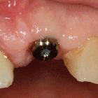
An implant
ready to be restored. The temporary healing abutment is visible in the mouth.
Dental implants are a common procedure used to replace absent teeth. Radiology has a role in pre-implant planning as well as post-implant assessment including identification of complications.
Radiographic features
Dental cone beam computed tomography (CBCT) is the most common modality used in dental implant imaging.
Pre-implant imaging
Assessment of the density (i.e. quality) and alveolar height and width (i.e. quantity) of bone is key for success implant osseointegration :
- density
- edentulism typically leads to bone loss with resorption of the alveolar process typically affecting width before height. Mandibular bone resorption is centrifugal whereas maxillary is centripetal.
- a number of Bone Quality Indices exist
- Cawood and Howell
- Lekholm and Zarb
- Norton and Gamble
- quantity
- bone implant site needs to be at least 7-9 mm high and 5 mm wide
- measurements should be taken every four sagittal oblique images through the region planned for implant
- other features
- contours
- dental extractions
- periodontal disease
- retained dental roots
- periapical lucencies
- anatomical variations, e.g. mandibular or maxillary tori
- pre-implant sinus lift or other procedure to increase maxillary alveolar ridge height and width
Siehe auch:
- Periimplantitis Zahnimplantat
- MRT bei Zahnimplantaten
- Artefakte durch Zahnimplantate
- Sinus-Lift-Prozedur
und weiter:


 Assoziationen und Differentialdiagnosen zu Zahnimplantate: