dorsal arachnoid web
Dorsal thoracic arachnoid web refers to a thickened band of arachnoid over the dorsal aspect of the cord. It usually causes a focal thoracic cord distortion with consequent neurological dysfunction.
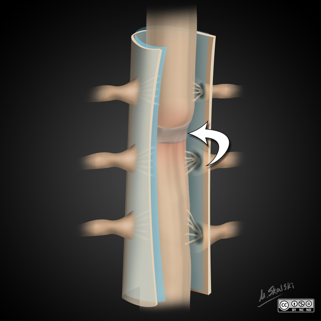
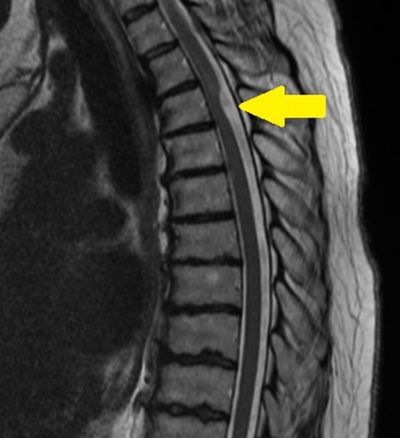
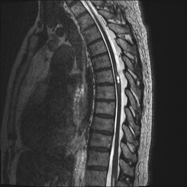
On imaging, it is characterized by a focal dorsal indentation and anterior displacement of the thoracic spinal cord leading to widening of the dorsal CSF space.
Clinical presentation
Due to the limited number of reported cases the incidence of this condition may well be under-recognized. The cases reported have a variety of signs and symptoms attributed to the band, including :
- episodic weakness and sensory symptoms, sometimes relieved by recumbency
- hyperreflexia, spastic paraparesis, clonus, and hypertonia
- pain
- gait instability
In most cases, there is no history of significant prior trauma or surgery .
Pathology
This condition is due to the presence of a thickened band of arachnoid over the dorsal aspect of the cord. This results in a focal displacement of the cord anteriorly and is often associated with syringomyelia, believed to be due to altered CSF flow dynamics due to the aforementioned web .
Radiographic features
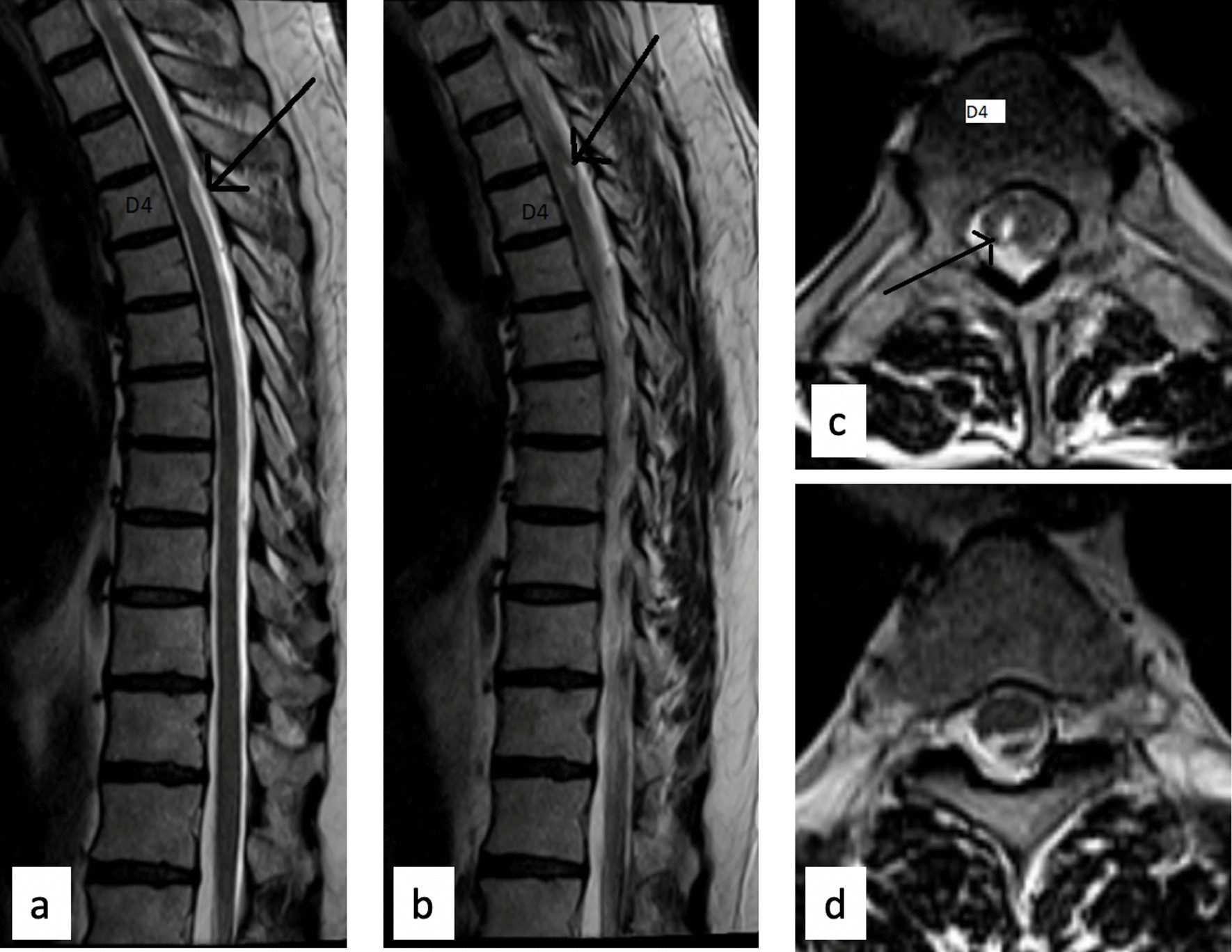
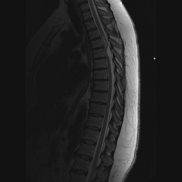
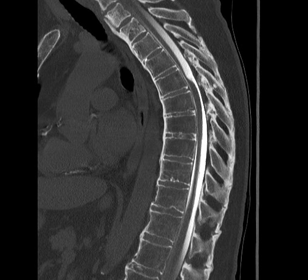
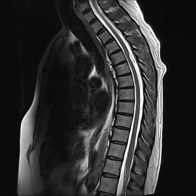
Although direct visualization of the web is beyond routine imaging; the key feature which implies the diagnosis is a focal dorsal indentation and anterior displacement of the thoracic cord, best identified on MRI (with CT myelogram as an alternative).
MRI
The thoracic cord appears focally displaced anteriorly, with the widening of the dorsal CSF space. The outline of this enlarged CSF space on sagittal imaging has been likened to the silhouette of a surgical scalpel and has been termed the scalpel sign .
The thoracic cord above or below the band often demonstrates a high T2 signal sometimes with a defined syrinx.
Treatment and prognosis
Provided the diagnosis is suspected, neurosurgical intervention with resection of the band can be curative .
Differential diagnosis
Considerations include
- ventral cord herniation
- cord pulled rather than pushed forward
- no space between cord and ventral theca (this may also be true of both arachnoid cysts and dorsal arachnoid webs, however)
- focal distortion at the point of herniation
- herniation may be visible
- dorsal spinal arachnoid cyst
- appearances are very similar, but cyst can be demonstrated on myelography (usually fill with contrast slower than the rest of the subarachnoid space)
- distortion of the cord is less focal, i.e. no-scalpel sign
Other causes of intramedullary cysts/abnormal signal should also be considered, and contrast is necessary to exclude an intramedullary mass (e.g. ependymoma).
Siehe auch:
und weiter:


 Assoziationen und Differentialdiagnosen zu intraspinales arachnoidales Web: