Duplikation der Hypophyse

Magnetic
resonance imaging of sellar and juxtasellar abnormalities in the paediatric population: an imaging review. Pituitary duplication. Coronal post-contrast images (a and b) show two pituitary stalks which extend inferiorly into separate duplicated pituitary glands. Sagittal T1WI (c) shows marked thickening of the hypothalamus, referred to as a pseudohamartoma, which has intermediate signal. The pseudohamartoma showed no contrast enhancement (not shown)
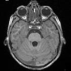
Duplication
of the pituitary gland. MRI Axial and Coronal T1 images showing complete duplication of the pituitary gland with two posterior bright spots
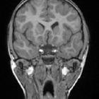
Duplication
of the pituitary gland. MRI Axial and Coronal T1 images showing complete duplication of the pituitary gland with two posterior bright spots
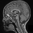
Duplication
of the pituitary gland. MRI Sagittal T1image showing thickening of the floor of the third ventricle (arrow) in keeping with fusion of the mamillary bodies and tuber cinerum
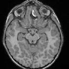
Duplication
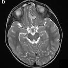
of the pituitary gland. MRI. a-b Axial T1 and T2 images showing anterior frontal mass which contains hyperintense T1 fatty tissue, c Axial SWI image showing chemical shift artefact as well as possible blooming artefact suggesting additional calcification
Duplication
of the pituitary gland. MRI. a-b Axial T1 and T2 images showing anterior frontal mass which contains hyperintense T1 fatty tissue, c Axial SWI image showing chemical shift artefact as well as possible blooming artefact suggesting additional calcification
Duplikation der Hypophyse
Siehe auch:

 Assoziationen und Differentialdiagnosen zu Duplikation der Hypophyse:

