DWI bei akuter Cholezystitis
Diffusion-weighted
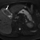
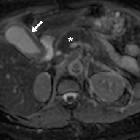
MRI: findings and role in acute cholecystitis. High (800) b-value diffusion-weighted images (d,e) showed visually hyperintense signal in the gallbladder wall (arrows) indicating acute inflammation, and pancreatic gland (*) gradually progressive hypersignal consistent with oedematous acute pancreatitis from tail to head.

Diffusion-weighted
MRI: findings and role in acute cholecystitis. High (800) b-value diffusion-weighted images (d,e) showed visually hyperintense signal in the gallbladder wall (arrows) indicating acute inflammation, and pancreatic gland (*) gradually progressive hypersignal consistent with oedematous acute pancreatitis from tail to head.
Diffusion-weighted
MRI: findings and role in acute cholecystitis. Apparent diffusion coefficient map showed hypointensity indicating restricted diffusion in gallbladder wall (arrow, 1.45-1.6x10⁻³ mm2/s) and pancreas (*, progressively increasing from 0.8 at head to 1.1x10⁻³ mm2/s in tail).


 Assoziationen und Differentialdiagnosen zu DWI bei akuter Cholezystitis: