eingeschlagenes Periost

Trapped
periosteum managed conservatively. Widened lateral physis of right femur.

Trapped
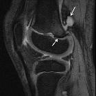
periosteum managed conservatively. Increased signal along physis of distal femur with surrounding oedema in metaphysis and adjacent epiphysis (lower left arrow). Fluid collection at posterior adjacent soft tissue represents periosteal and capsular injury (upper right arrow).

Trapped
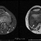
periosteum managed conservatively. Prominent low signal region at physis (arrow) indicates trapped periosteum.

Trapped
periosteum managed conservatively. Proton density imaging highlights adjacent bone marrow oedema. Low prominent signal is again appreciated indicating trapped periosteum (arrow).

Trapped
periosteum managed conservatively. A segment of missing periosteum is seen at the posterior lateral aspect of the distal femur near the metaphysis.

Trapped
periosteum managed conservatively. A few slices more inferiorly a well trapped periosteum is seen.
eingeschlagenes Periost
Siehe auch:

 Assoziationen und Differentialdiagnosen zu eingeschlagenes Periost:
