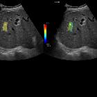
Elastographie

Epididymal
haematoma: multiparametric ultrasonographic findings.. Colour-coded elastographic image shows the lesion to be predominantly homogeneously soft except for the part where multiple colours can be seen. (Blue colour represents soft and red colour represents hard tissue).

Elastography
• Sonoelastography of cervical lymph nodes - Ganzer Fall bei Radiopaedia

Cirrhosis •
Cirrhosis (elastography) - Ganzer Fall bei Radiopaedia

Elastography
• Fibroadenoma - Ganzer Fall bei Radiopaedia

Elastography
• Invasive micropapillary carcinoma of the breast - Ganzer Fall bei Radiopaedia

Elastography
• Infiltrating pleiomorphic lobar carcinoma - Ganzer Fall bei Radiopaedia

Tubular
adenoma of the breast • Tubular adenoma - breast - Ganzer Fall bei Radiopaedia
Elastography is a newer technique that exploits the fact that a pathological process alters the elastic properties of the involved tissue. This change in elasticity is detected and imaged using elastography.
Radiographic technique
Sono-elastography
Sono-elastography is the term used when ultrasound is used to assess elastography
- strain elastography (also known as static or compression elastography)
- shear wave elastography (also known as transient elastography)
MR elastography
MR elastography is the term used when MRI is used to assess tissue stiffness. It uses shear waves to assess the tissue displacement in all directions making it more precise than sonoelastography.
MR elastography has been most widely used in cases of liver fibrosis where larger lesions can be easily assessed even in the presence of ascites.
Uses
- differentiating malignant and benign neoplasms (especially breast)
- identifying early traumatic changes in muscles and tendons
- aiding in deciding the biopsy site more accurately, reducing negative biopsy rates
- assessing liver fibrosis
- assessing liver steatosis (eg non-alcoholic fatty liver disease and steatohepatitis)
Siehe auch:
und weiter:

