Embolisationen Arteria renalis und Niere

Endovascular
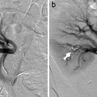
intervention in renovascular disease: a pictorial review. Right RAVM as seen on CT (a, white arrow) and angiogram (b, black arrow). Coil embolisation provides an effective option in the treatment of larger RAVMS (c, black arrow)

Endovascular
intervention in renovascular disease: a pictorial review. Angiographic image of a right RAVF (a, black arrow) developed following ultrasound-guided biopsy of a transplant kidney with subsequent selective coil embolisation of arterial feeders (b, white arrow)

Endovascular
intervention in renovascular disease: a pictorial review. A large right RCC as demonstrated on CT (a, long white arrow) and angiographic imaging (b, black arrow). In this case, renal artery TCE (c, white arrows) allowed for tumour devascularisation prior to surgical resection

Endovascular
intervention in renovascular disease: a pictorial review. Large left renal AML as seen on CT (a, white arrow) and angiography (b, black arrow) subsequently embolised with sclerosing agent (ethanol) (c)

Endovascular
intervention in renovascular disease: a pictorial review. Right renal trauma with persistent haemorrhage of an interlobar artery following a cycling accident demonstrated by angiography (a, white arrow). Therapeutic management consisted of coil embolisation of the injured vessel (b, black arrow)

Endovascular
intervention in renovascular disease: a pictorial review. A true extraparenchymal saccular right renal artery aneurysm seen on MR (a, long white arrow) and angiography (b, black arrow) effectively managed with intraaneurysmal coil embolisation (c, white arrow)

Endovascular
intervention in renovascular disease: a pictorial review. Axial CT (a, long white arrow) and angiographic (b, black arrow) images of a left intraparenchymal renal artery pseudoaneurysm subsequent to partial nephrectomy for renal tumour. The propensity for severe haemorrhage with these lesions warranted selective coil embolisation (c, white arrow)

Renal
arteriovenous fistula: Which US findings are expected?. After using coils (green arrow), blood keeps flowing through the arteriovenous fistula (red arrow).

Renal
arteriovenous fistula: Which US findings are expected?. After using more coils (yellow arrow), blood flow could not be interrupted (red arrow).

Renal
arteriovenous fistula: Which US findings are expected?. A non-adhesive liquid embolic agent (white arrows) was required to interrupt the blood flow within the fistula (red arrow).
Embolisationen Arteria renalis und Niere
Siehe auch:
- arteriovenöse Fistel der Niere
- Embolisation Angiomyolipom der Niere
- Nierentumorembolisation
- Embolisation Aneurysma spurium der Arteria renalis
- Embolisation arteriovenöse Fistel der Niere
- Embolisation Nierenruptur
- Embolisation arteriovenöse Malformation der Niere
- palliative Nierentumorembolisation
- endovaskuläre Behandlung Nierenarterienaneurysma

 Assoziationen und Differentialdiagnosen zu Embolisationen Arteria renalis und Niere: