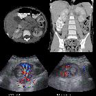
Endometriose der Niere

Laparoscopic
Resection of Renal Capsular Endometriosis in a Woman with Menstrual-Related Flank Pain: Case Report. a Abdominopelvic CT showing a heterogeneous mass (black arrow) with pressure effect on the upper pole of the right kidney (coronal view). b. Abdominopelvic CT showing a heterogeneous mass (black arrow) with pressure effect on the upper pole of the right kidney (axial view)

Laparoscopic
Resection of Renal Capsular Endometriosis in a Woman with Menstrual-Related Flank Pain: Case Report. Abdominopelvic CT showing malrotated right kidney (black star) and anteriorly oriented right renal pelvis

Endometriosis
in a kidney with focal xanthogranulomatous pyelonephritis and a perinephric abscess. Contrast computerized tomography images. It shows the replacement of the right renal tissue by several rounded, low density areas. A perinephric abscess is noted invading the right psoas muscle (arrows). Renal stones, the frequent etiology of xanthogranulomatous pyelonephritis, are also seen. a Transverse section. b Coronal section
Endometriose der Niere
Siehe auch:

 Assoziationen und Differentialdiagnosen zu Endometriose der Niere:


